. 2017 Feb;5(1):37-44.
doi: 10.1177/2050640616644142. Epub 2016 Jun 23.

Distinct metaplastic and inflammatory phenotypes in autoimmune and adenocarcinoma-associated chronic atrophic gastritis

Affiliations

Distinct metaplastic and inflammatory phenotypes in autoimmune and adenocarcinoma-associated chronic atrophic gastritis

Sangho Jeong et al. United European Gastroenterol J. 2017 Feb.

Abstract

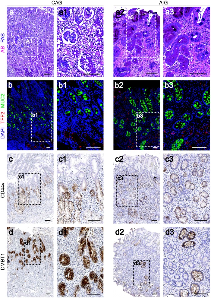
Background: Autoimmune gastritis (AIG) and adenocarcinoma-associated chronic atrophic gastritis (CAG) are both associated with oxyntic atrophy, but AIG patients demonstrate an increased risk of carcinoid tumors rather than the elevated risk of adenocarcinoma observed with CAG. We therefore sought to compare the characteristics of the metaplastic mucosa in AIG and CAG patients.

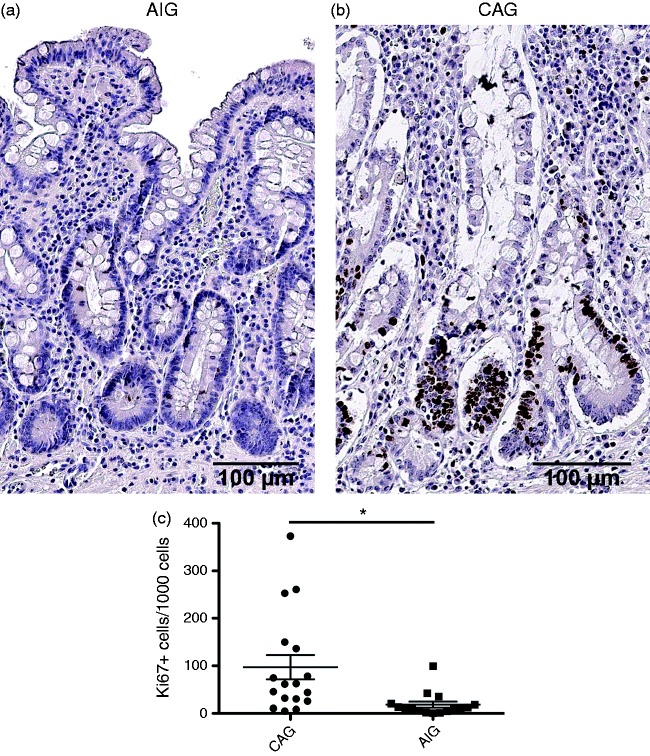
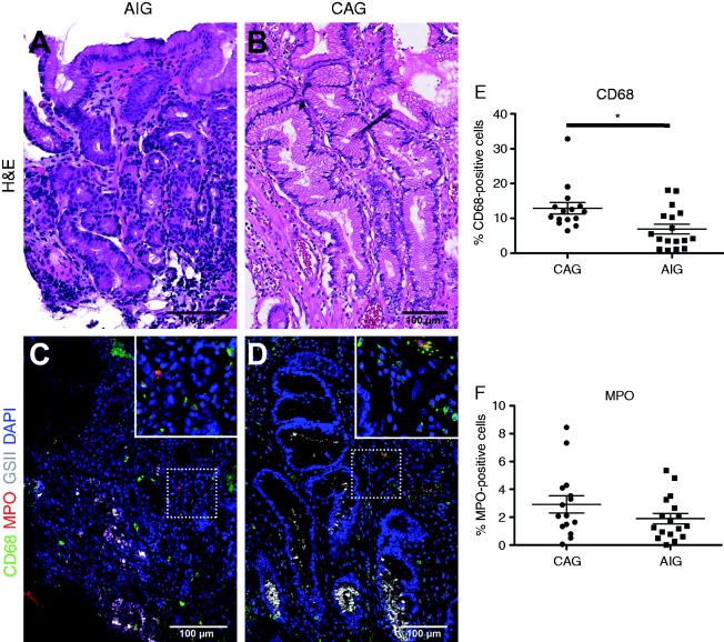
Methods: We examined markers for metaplasia (spasmolytic polypeptide expressing metaplasia (SPEM) and intestinal metaplasia) as well as proliferation (Ki67) and immune cell populations (neutrophils, macrophages, and eosinophils) in gastric sections from 16 female patients with autoimmune thyroiditis and AIG and 17 patients with CAG associated with gastric adenocarcinoma.

Results: Both AIG and CAG patients demonstrated prominent SPEM and intestinal metaplasia. However, AIG patients displayed significantly lower numbers of infiltrating macrophages and significantly reduced mucosal cell proliferation as compared to CAG patients.

Conclusions: These findings indicate that, while both AIG and CAG patients display prominent oxyntic atrophy and metaplasia, the AIG patients do not show proliferative metaplastic lineages that would predispose to adenocarcinoma.

Keywords: CD44 variant 9; DMBT1; Spasmolytic polypeptide-expressing metaplasia; gastritis; intestinal metaplasia; macrophage; neutrophil.


Figures

Figure 1.
Figure 1.
Metaplasia in the gastric mucosa of autoimmune gastritis (AIG) and chronic atrophic gastritis (CAG) patients. Top row: dual Alcian blue and periodic acid-Schiff (AB/PAS) staining of gastric mucosa in CAG ((a), (a1)) and AIG ((a2), (a3)) patients. Note AB staining of intestinal metaplasia in luminal cells, and PAS-staining of spasmolytic polypeptide expressing metaplasia (SPEM) at the bases of the gland. Second row: dual immunofluorescence staining of TFF2 (Trefoil Factor 2, SPEM marker, red) and MUC2 (Mucin 2, intestinal metaplasia marker, green) in gastric mucosa from CAG ((b), (b1)) and AIG ((b2), (b3)). In all cases nuclei were labeled with 4′,6-diamidino-2-phenylindole (DAPI) (blue). Images (a1), (a3), (b1) and (b3) are higher magnification as noted by white boxed outlines. Third row: immunohistochemical staining of CD44v (SPEM marker) in CAG ((c), (c1)) and AIG ((c2), (c3)) gastric mucosa. Bottom row: immunohistochemical staining of DMBT1 (Deleted in malignant brain tumors 1, intestinal metaplasia marker) for CAG ((d), (d1)), and AIG ((d2), (d3)) mucosa. Images (c1), (c3), (d1) and (d3) are higher magnification as noted by black-boxed outlines. Bar indicates 100 µm.
Figure 2.
Figure 2.
Characteristics of proliferation in metaplastic mucosa. Representative immunostaining for Ki67 in (a) a chronic atrophic gastritis (CAG) patient and (b) an autoimmune gastritis (AIG) patient. (c) Quantitation of Ki67-positive cells demonstrates a markedly lower level of proliferation in AIG patients. In CAG patients, filled circles indicate females and open circles indicate males. *p = 0.0066. Bar indicates 100 µm.
Figure 3.
Figure 3.
Characteristics of inflammatory infiltrates in metaplastic mucosa. Comparison of the infiltration of macrophages (CD68, green) and neutrophils (MPO, Myeloperoxidase, red) between chronic atrophic gastritis (CAG) ((a), (c)) and autoimmune gastritis (AIG) ((b), (d)) patients. Hematoxylin and eosin (H&E) staining of sections are shown from AIG (a) and CAG (b) patient stomach. In (c) and (d), spasmolytic polypeptide expressing metaplasia (SPEM) cells were stained with Griffonia simplicifolia II-lectin (white) and nuclei were stained with 4′,6-diamidino-2-phenylindole (DAPI) (blue). Insets show higher magnification images as noted by white boxed outlines. Bar indicates 100 µm. (e) Quantitation of immunostaining demonstrated the presence of significantly fewer CD68-positive macrophages in AIG patients as compared to CAG patients (*p < 0.01).

References

    1. Nomura S, Baxter S, Yamaguchi T, et al. Spasmolytic polypeptide expressing metaplasia (SPEM) to pre-neoplasia in H. felis-infected mice. Gastroenterology 2004; 127: 582–594. - PubMed
    1. Correa P. A human model of gastric carcinogenesis. Cancer Res 1988; 48: 3554–3560. - PubMed
    1. Goldenring JR, Nam KT, Wang TC, et al. Spasmolytic polypeptide-expressing metaplasia and intestinal metaplasia: Time for reevaluation of metaplasias and the origins of gastric cancer. Gastroenterology 2010; 138: 2207–2210. - PMC - PubMed
    1. Nam KT, Lee H-J, Sousa JF, et al. Mature chief cells are cryptic progenitors for metaplasia in the stomach. Gastroenterology 2010; 139: 2028–2037. - PMC - PubMed
    1. Weis VG, Sousa JF, LaFleur BJ, et al. Heterogeneity in mouse SPEM lineages identifies markers of metaplastic progression. Gut 2013; 62: 1270–1279. - PMC - PubMed