Rare Clinical Sign of Hodgkin's Lymphoma: Ocular Involvement
- PMID: 28405486
- PMCID: PMC5384116
- DOI: 10.4274/tjo.92609
Rare Clinical Sign of Hodgkin's Lymphoma: Ocular Involvement
Abstract
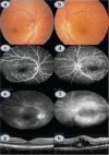
Bilateral non-granulomatous anterior uveitis with left vitritis and macular edema were detected in a 19-year-old woman presenting with blurred vision in her left eye. Light microscopic study of the pathologic mediastinal lymph node that was detected via contrast computed tomography imaging during etiologic study revealed nodular sclerosing and mixed cellularity Hodgkin's lymphoma (HL). Ocular findings completely resolved with adriablastin, bleomycin, vinblastine, dacarbazine chemotherapy treatment. Herein, it is emphasized that HL should be remembered as one of the differential diagnoses in patients with ocular inflammatory pathologies such as uveitis and vasculitis. The ocular findings of HL are discussed.
Keywords: Anterior uveitis; Hodgkin’s lymphoma; Macular edema; posterior uveitis.
Conflict of interest statement
Conflict of Interest: No conflict of interest was declared by the authors.
Figures


References
-
- Knowles MD. Neoplastic hematopathology (2nd ed) New York: Lippincott Williams &Wilkins; 2001. pp. 610–680.
-
- Young GA. Lymphoma at uncommon sites. Hematol Oncol. 1999;17:53–83. - PubMed
-
- Şimşek HC, Akkoyun İ, Yılmaz G. Hematolojik Hastalıklarda Göz Bulguları. Retina-Vitreus. 2014;22:85–92.
-
- Mosteller MW, Margo CE, Hesse RJ. Hodgkin’s disease and granulomatous uveitis. Ann Ophthalmol. 1985;17:787–790. - PubMed
-
- Primbs GB, Monsees WE, Irvine AR., Jr Intraocular Hodgkin’s disease. Arch Ophthalmol. 1961;66:477–482. - PubMed
LinkOut - more resources
Full Text Sources
Other Literature Sources
