Arteriovenous Malformation Underlying a Plexiform Neurofibroma: An Unusual Presentation
- PMID: 28405554
- PMCID: PMC5372434
- DOI: 10.4103/2229-5178.202272
Arteriovenous Malformation Underlying a Plexiform Neurofibroma: An Unusual Presentation
Abstract
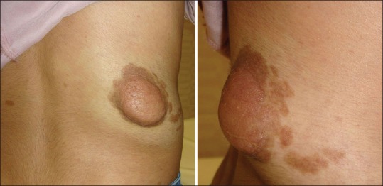
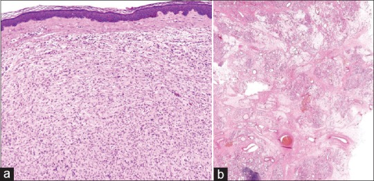
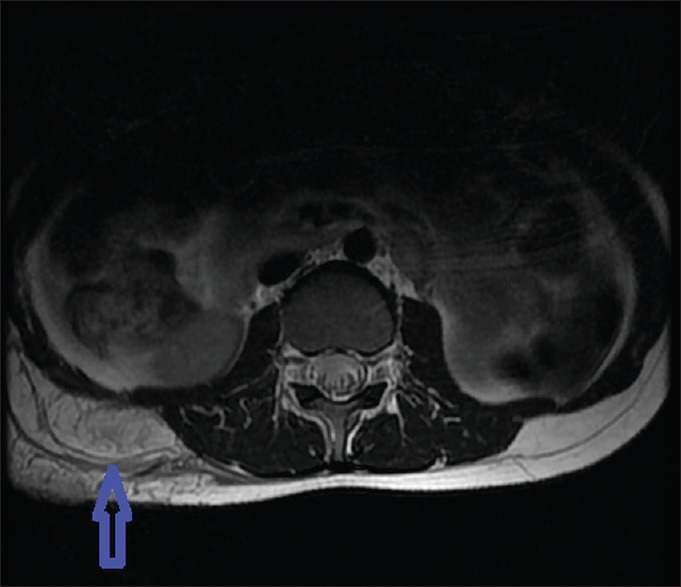
Vascular abnormalities associated with neurofibromatosis type 1 are well described in the literature, however, arteriovenous malformation is a very rare finding in neurofibromatosis type 1. We report the case of an 11-year-old girl who presented with a soft mass on the right flank. Provisional diagnosis of plexiform neurofibroma was made on the basis of clinical and histological observations. Because the lesion was warm on palpation, imaging studies were performed to evaluate further and arteriovenous malformation was detected underlying the plexiform neurofibroma. This report emphasizes the importance of careful examination and proper investigations of the plexiform neurofibroma prior to treatment strategies to avoid future complications. The rarity of plexiform neurofibroma in association with arteriovenous malformation at the same site was also highlighted in this report.
Keywords: Arteriovenous malformation; hypervascularity; neurofibromatosis; plexiform.
Conflict of interest statement
There are no conflicts of interest.
Figures
Similar articles
-
Facial plexiform neurofibroma in a child with neurofibromatosis type I: a case report.J Indian Soc Pedod Prev Dent. 2007 Mar;25(1):30-5. doi: 10.4103/0970-4388.31987. J Indian Soc Pedod Prev Dent. 2007. PMID: 17456965
-
Unexpected diagnosis of superficial neurofibroma in a lesion with imaging features of a vascular malformation.Pediatr Radiol. 2005 Dec;35(12):1250-3. doi: 10.1007/s00247-005-1565-9. Epub 2005 Sep 9. Pediatr Radiol. 2005. PMID: 16151788
-
The recurrent plexiform neurofibroma of the scalp in neurofibromatosis type 1: illustrative case.J Neurosurg Case Lessons. 2021 Jan 11;1(2):CASE2024. doi: 10.3171/CASE2024. eCollection 2021 Jan 11. J Neurosurg Case Lessons. 2021. PMID: 35854934 Free PMC article.
-
Diffuse ganglioneuromatosis and plexiform neurofibroma of the urinary bladder: report of a pediatric example and literature review.Hum Pathol. 2008 Nov;39(11):1708-12. doi: 10.1016/j.humpath.2008.02.019. Epub 2008 Jul 24. Hum Pathol. 2008. PMID: 18656232 Review.
-
Congenital extra calvarial plexiform neurofibroma in occipito-cervical region with Occipital bone defect with neurofibromatosis type 1 and segmental neurofibromatosis: Case report and review of literature.J Pediatr Neurosci. 2016 Oct-Dec;11(4):295-297. doi: 10.4103/1817-1745.199469. J Pediatr Neurosci. 2016. PMID: 28217149 Free PMC article. Review.
References
-
- Lin AE, Birch P, Korf BR, Tenconi R, Niimura M, Poyhonen M, et al. Cardiovascular malformations and other cardiovascular abnormalities in Neurofibromatosis type I. Am J Med Genet. 2000;95:108–17. - PubMed
-
- Hamilton SJ, Friedman JM. Insights into the pathogenesis of neurofibromatosis 1 vasculopathy. Clin Genet. 2000;58:341–4. - PubMed
-
- Gottfried ON, Viskochil DH, Couldwell WT. Neurofibromatosis Type 1 and tumorigenesis: Molecular mechanisms and therapeutic implications. Neurosurg Focus. 2010;28:8. - PubMed
Publication types
LinkOut - more resources
Full Text Sources
Other Literature Sources
Research Materials