Comparative Study
. 2017 Apr 6;2(7):e90443.
doi: 10.1172/jci.insight.90443.

MicroRNA-125a and -b inhibit A20 and MAVS to promote inflammation and impair antiviral response in COPD

Affiliations
Comparative Study

MicroRNA-125a and -b inhibit A20 and MAVS to promote inflammation and impair antiviral response in COPD

Alan C-Y Hsu et al. JCI Insight. .

Abstract

Influenza A virus (IAV) infections lead to severe inflammation in the airways. Patients with chronic obstructive pulmonary disease (COPD) characteristically have exaggerated airway inflammation and are more susceptible to infections with severe symptoms and increased mortality. The mechanisms that control inflammation during IAV infection and the mechanisms of immune dysregulation in COPD are unclear. We found that IAV infections lead to increased inflammatory and antiviral responses in primary bronchial epithelial cells (pBECs) from healthy nonsmoking and smoking subjects. In pBECs from COPD patients, infections resulted in exaggerated inflammatory but deficient antiviral responses. A20 is an important negative regulator of NF-κB-mediated inflammatory but not antiviral responses, and A20 expression was reduced in COPD. IAV infection increased the expression of miR-125a or -b, which directly reduced the expression of A20 and mitochondrial antiviral signaling (MAVS), and caused exaggerated inflammation and impaired antiviral responses. These events were replicated in vivo in a mouse model of experimental COPD. Thus, miR-125a or -b and A20 may be targeted therapeutically to inhibit excessive inflammatory responses and enhance antiviral immunity in IAV infections and in COPD.


Conflict of interest statement

Conflict of interest: The authors have declared that no conflict of interest exists.

Figures
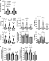
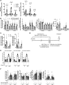
Figure 1
Figure 1. IAV infection is more severe and results in exaggerated inflammatory but impaired antiviral responses in pBECs from patients with COPD.
pBECs from healthy controls, COPD patients, and healthy smokers were infected with human IAV H3N2 or H1N1, and (A) virus replication was measured at 24 hours. (B) Proinflammatory cytokines/chemokines IL-6, CXCL-8, TNF-α, and IL-1β and antiviral cytokines IFN-β and IFN-λ1 were measured in culture supernatants at 24 hours. (C) Phospho-p65 was assessed at 6 hours and 24 hours, and densitometry results (from Supplemental Figure 1A, representative immunoblot) were calculated as phospho-p65/GAPDH ratios and expressed as fold change from healthy media control. Data are mean ± SEM, n = 15 (healthy controls and COPD patients) or 5 (healthy smokers). *P ≤ 0.05 versus respective uninfected media control, +P ≤ 0.05 versus infected or uninfected healthy controls. Statistical differences were determined with one-way ANOVA followed by Bonferroni post-test.
Figure 2
Figure 2. IAV infection is more severe and results in exaggerated inflammatory and impaired antiviral responses in experimental COPD.
(A) BALB/c mice were exposed to cigarette smoke (Smk) or normal air (Air) for 8 weeks, infected with IAV H1N1 (A/PR/8/34, 8 pfu) or media (Sham) on the last day of smoke exposure, and sacrificed 7 days postinfection (dpi). (B) Virus titers were measured in bronchoalveolar lavage fluid. (C) IL-6, KC, TNF-α, and IL-1β and (D) IFN-β and IFN-λ3 were assessed in lung homogenates. (E) Phospho-p65 protein was determined in lung homogenates. Densitometry results (from Supplemental Figure 1D, representative immunoblot) were calculated as phospho-p65 or IFN-β/β-actin ratios and expressed as fold change from Air sham control. Data are mean ± SEM, n = 6–8 per group. *P ≤ 0.05 versus Sham control, +P ≤ 0.05 versus Air control. Statistical differences were determined with one-way ANOVA followed by Bonferroni post-test.
Figure 3
Figure 3. A20 expression is reduced and negatively regulates inflammatory but not antiviral responses in pBECs from patients with COPD.
(A) pBECs were infected with human IAV H3N2 or H1N1, and the protein levels of A20 were determined at 6 hours and 24 hours. Densitometry results (from Supplemental Figure 2A, representative immunoblot) were calculated as A20 or phospho-p65/GAPDH ratios and expressed as fold change from healthy media control. Data are mean ± SEM, n = 15 per group. *P ≤ 0.05 versus respective uninfected media control, +P ≤ 0.05 versus healthy control. A20 expression was inhibited with a specific siRNA, pBECs were infected with IAVs, and protein levels of (B) A20; (C) phospho-p65; (D) cytokines/chemokines IL-6, CXCL-8, TNF-α, and IL-1β; and antiviral (E) IFN-β and IFN-λ1 were measured 24 hours later. Densitometric ratios (from Supplemental Figure 2C, representative immunoblot) were expressed as fold change from untreated media control. Data are mean ± SEM, n = 3 per group. *P ≤ 0.05 versus untreated, uninfected media control, +P ≤ 0.05 versus untreated infected or uninfected control. Statistical differences were determined with one-way ANOVA followed by Bonferroni post-test.
Figure 4
Figure 4. IAV infection increases the levels of miR-125 and -b that suppress the production of A20, increase inflammatory responses, and reduce antiviral responses in human COPD pBECs and experimental COPD.
pBECs were infected with human IAV H3N2 or H1N1, and (A) miR-125a and -b levels were assessed at 24 hours. Data are mean ± SEM, n = 15 per group. *P ≤ 0.05 versus uninfected media control, +P ≤ 0.05 versus healthy or smoker control. (B) pBECs were treated with miR-125a or -b antagomir and were infected, and the levels of A20, phospho-p65, IFN-β, and IFN-λ1 were assessed. Densitometry results (Supplemental Figure 3B, representative immunoblot) were calculated as A20 or phospho-p65/GAPDH ratios and expressed as fold change from untreated, uninfected control. Data are mean ± SEM, n = 3 per group. *P ≤ 0.05 versus untreated, uninfected media control; +P ≤ 0.05 versus untreated infected or uninfected group. (C) BALB/c mice were exposed to cigarette smoke (Smk) or normal air (Air) for 8 weeks, inoculated with IAV H1N1 (A/PR/8/34, 8 pfu) or media (Sham) on the last day of smoke exposure, and sacrificed 7 days postinfection (dpi). The levels of miR-125a and -b were measured. Data are mean ± SEM, n = 6–8 per group. *P ≤ 0.05 versus Sham group, +P ≤ 0.05 versus Air-infected or uninfected group. (D) In other groups, on the last day of smoke exposure, mice were treated with miR-125a or -b antagomir alone or in combination and infected with IAV, and (E) airway histological scores were assessed. Data are mean ± SEM, n = 6–8 per group. *P ≤ 0.05 versus infected and scrambled treated Air controls, +P ≤ 0.05 versus infected and scramble-treated Smk group. (F) The protein levels of A20, phospho-p65, and IFN-β in lung homogenates were also measured. Densitometry results (Supplemental Figure 4D) were calculated as A20 or phospho-p65/β-actin ratios and expressed as fold change from untreated, uninfected control. Data are mean ± SEM, n = 6–8 per group. *P ≤ 0.05 versus infected scrambled-treated Air group, +P ≤ 0.05 versus infected scrambled Smk group. Statistical differences were determined with one-way ANOVA followed by Bonferroni post-test.
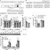
Figure 5
Figure 5. miR-125a and -b target a functional binding site of the 3′-UTR of the mRNA of MAVS to suppress its expression.
(A) Representation of MAVS gene structure and location of miR-125a and -b binding site. (B) The binding site on 3′-UTR of MAVS is 100% conserved between human and mouse MAVS. (C) pBECs were infected with H3N2 or H1N1, and MAVS protein was detected at 6 hours (left) and 24 hours (right). Densitometry results (Supplemental Figure 5A, representative immunoblot) were calculated as MAVS/GAPDH ratios and expressed as fold change from untreated, uninfected controls. Data are mean ± SEM, n = 15 per group. *P ≤ 0.05 versus uninfected healthy or smoker controls, +P ≤ 0.05 versus infected or uninfected healthy controls. (D) BALB/c mice were exposed to cigarette smoke (Smk) or normal air (Air) for 8 weeks, inoculated with IAV H1N1 (A/PR/8/34, 8 pfu) or media (Sham) on the last day of smoke exposure, and sacrificed 7 days postinoculation (dpi). The levels of MAVS protein were measured in lung homogenates. Densitometry results (Supplemental Figure 5B, representative immunoblot) were calculated as MAVS/β-actin ratios in mouse and expressed as fold change from untreated, uninfected controls. Data are mean ± SEM, n = 6 per group. *P ≤ 0.05 versus Sham-treated controls, +P ≤ 0.05 versus infected Air controls. (E) The miR-125a and -b binding site on 3′-UTR was cloned into a pMIR luciferase reporter construct and transfected into HEK293 cells with miR-125a or -b mimetics. The luciferase reporter assay was performed to determine binding. Data are mean ± SEM, n = 3 per group.*P ≤ 0.05 versus miRNA scrambled controls. (F) Ago2 was immunoprecipitated from miR-125a or -b mimetic-transfected HEK293, and (G) A20 and MAVS mRNA was detected by qPCR in Ago2-immunoprecipitate. Data are mean ± SEM, n = 3 per group. *P ≤ 0.05 versus IgG control IP. Statistical differences were determined with one-way ANOVA followed by Bonferroni post-test.
Figure 6
Figure 6. miR-125a and -b suppress the induction of MAVS and promote virus replication in human COPD pBECs and experimental COPD.
(A) miR-125a and -b antagomir or mimetics were added to pBECs before infection with human IAV H3N2 or H1N1, and mitochondrial antiviral signaling (MAVS) protein were assessed 24 hours after infection. Densitometry results (Supplemental Figure 6A, representative immunoblot) were calculated as MAVS/GAPDH ratios and expressed as fold change from untreated, uninfected controls. Data are mean ± SEM, n = 3. *P ≤ 0.05 versus untreated, uninfected media controls; +P ≤ 0.05 versus untreated, infected or uninfected controls. (B) Virus replication was also measured. Data are mean ± SEM, n = 3. *P ≤ 0.05 versus untreated, infected controls. (C) BALB/c mice were exposed to cigarette smoke (Smk) or normal air (Air) for 8 weeks, treated with mir125a and/or -b antagomir, infected with IAV H1N1 (A/PR/8/34, 8 pfu) or media (Sham) on the last day of smoke exposure, and sacrificed 7 days postinoculation (dpi). MAVS protein was measured. Densitometry results (Supplemental Figure 6C, representative immunoblot) were calculated as MAVS/β-actin ratios and expressed as fold change from untreated, uninfected controls. Data are mean ± SEM, n = 6. *P ≤ 0.05 versus infected, scramble treated Air or Smk controls. (D) Virus replication was assessed. Data are mean ± SEM, n = 6. *P ≤ 0.05 versus infected, scramble-treated controls. Statistical differences were determined with one-way ANOVA followed by Bonferroni post-test.
Figure 7
Figure 7. Roles of miR-125a and -b in the regulation of inflammatory and antiviral responses in IAV infection.
Increased levels of miR-125a and -b, for example in COPD, reduce the protein expression of A20 that results in uncontrolled NF-κB activation, leading to exaggerated induction of proinflammatory cytokines. miR-125a and -b also target and reduce MAVS and antiviral type I and III IFN production. Inhibition of miR-125a and -b enhances MAVS and antiviral responses, and it suppresses viral infection.

References

    1. Hallstrand TS, Hackett TL, Altemeier WA, Matute-Bello G, Hansbro PM, Knight DA. Airway epithelial regulation of pulmonary immune homeostasis and inflammation. Clin Immunol. 2014;151(1):1–15. doi: 10.1016/j.clim.2013.12.003. - DOI - PubMed
    1. Hsu AC, See HV, Hansbro PM, Wark PA. Innate immunity to influenza in chronic airways diseases. Respirology. 2012;17(8):1166–1175. doi: 10.1111/j.1440-1843.2012.02200.x. - DOI - PubMed
    1. Chaouat A, et al. Role for interleukin-6 in COPD-related pulmonary hypertension. Chest. 2009;136(3):678–687. doi: 10.1378/chest.08-2420. - DOI - PubMed
    1. Meylan E, et al. RIP1 is an essential mediator of Toll-like receptor 3-induced NF-kappa B activation. Nat Immunol. 2004;5(5):503–507. doi: 10.1038/ni1061. - DOI - PubMed
    1. Monks BG, Martell BA, Buras JA, Fenton MJ. An upstream protein interacts with a distinct protein that binds to the cap site of the human interleukin 1 beta gene. Mol Immunol. 1994;31(2):139–151. doi: 10.1016/0161-5890(94)90086-8. - DOI - PubMed

Publication types

MeSH terms