

A rotating cerium anode X-ray system allows visualization of intramural coronary vessels after cardiac stem cell therapy for myocardial infarction
- PMID: 28405805
- PMCID: PMC10717793
- DOI: 10.1007/s12576-017-0537-9
A rotating cerium anode X-ray system allows visualization of intramural coronary vessels after cardiac stem cell therapy for myocardial infarction
Abstract
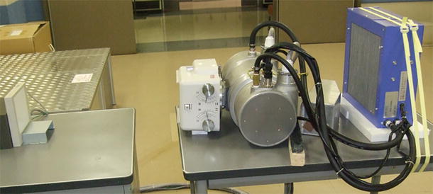
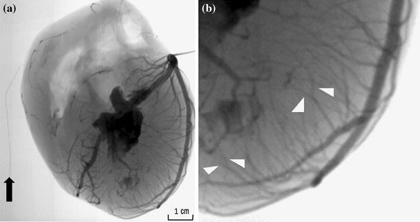
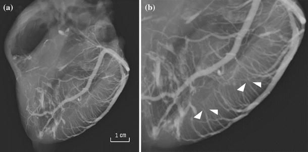
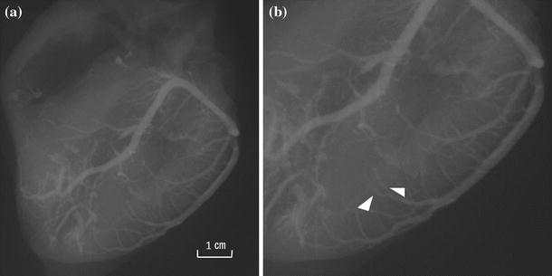
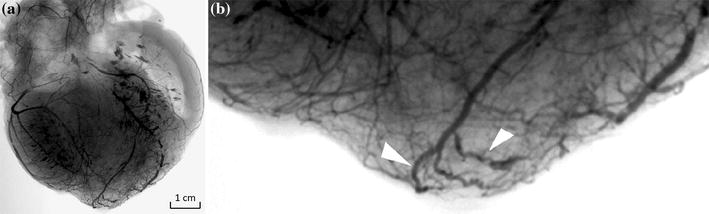
Conventional angiography is insufficient for evaluating the therapeutic effect of cardiac regeneration therapy. A microangiographic X-ray system using a cerium anode was developed. Cerium has a characteristic X-ray with a peak at 34.6 keV, which allows visualization of tiny amounts of iodine. The performance of the cerium anode X-ray system was evaluated in two excised normal canine hearts and in excised ischemic canine hearts treated with c-kit-positive cardiac stem cells (5 canines) or without cells (5 control canines). In the normal canines, branches penetrating from the left anterior descending artery into the myocardium were visualized, down to third-order branches. In just the treated hearts treated with stem cells, small vessels characterized by irregular vessel walls were observed. The cerium anode X-ray system allowed visualization of microvessels in excised ischemic canine hearts, and may evaluate the effect of cardiac stem cell therapy.
Keywords: C-kit-positive cardiac stem cells; Canine models; Cerium anode; Microangiography; Myocardial infarction.
Conflict of interest statement
The authors declare that they have no conflict interest other than the funding mentioned above.
Figures


References
-
- Borisenko O, Wylie G, Payne J, Bjessmo S, Smith J, Firmin R, Yonan N. The cost impact of short-term ventricular assist devices and extracorporeal life support systems therapies on the National Health Service in the UK. Interact Cardiovasc Thorac Surg. 2014;19:41–48. doi: 10.1093/icvts/ivu078. - DOI - PubMed
-
- Khazanie P, Hammill BG, Patel CB, Eapen ZJ, Peterson ED, Rogers JG, Milano CA, Curtis LH, Hernandez AF. Trends in the use and outcomes of ventricular assist devices among medicare beneficiaries, 2006 through 2011. J Am Coll Cardiol. 2014;63:1395–1404. doi: 10.1016/j.jacc.2013.12.020. - DOI - PMC - PubMed
-
- Rosalinda M, Linda WVL, Sean MD, Felix BE, Derek JH, Sandrine L, Jonathan L, Cinzia P, Rainer S, Kirsti Y, Ulf L, Christine LM, Stefan J, James W, Thomas E, Péter F, Joost PGS. Position paper of the european society of cardiology working group cellular biology of the heart: cell-based therapies for myocardial repair and regeneration in ischemic heart disease and heart failure. Eur Heart J. 2016 doi: 10.1093/eurheartj/ehw113. - DOI - PMC - PubMed
MeSH terms
Substances
Grants and funding
- 20390336/Grants-in-Aid for Scientific Research B
- 25861233/Grant-in-Aid for Young Scientists B
- H24-06-01/Grants-in-Aid from The Cardiovascular Research Fund, Tokyo, Japan
- 200624005A/Health and Labour Sciences Research Grant
- 22-5/The Research Funding for Longevity Sciences from National Center for Geriatrics and Gerontology
LinkOut - more resources
Full Text Sources
Other Literature Sources
Medical
