Review
. 2017 Apr 13;8(2):14.
doi: 10.3390/jfb8020014.

Artificial Red Blood Cells as Potential Photosensitizers in Dye Laser Treatment Against Port-Wine Stains

Affiliations
Review

Artificial Red Blood Cells as Potential Photosensitizers in Dye Laser Treatment Against Port-Wine Stains

Naoaki Rikihisa et al. J Funct Biomater. .

Abstract

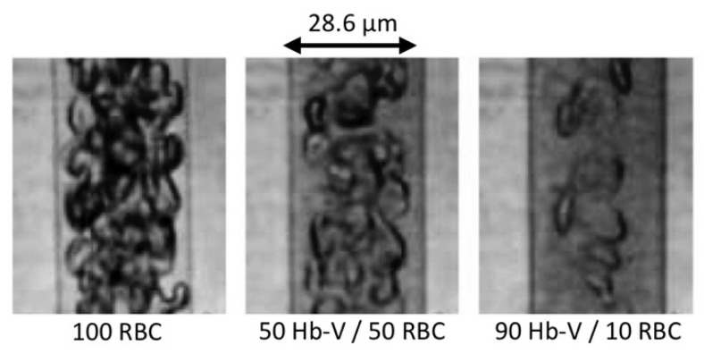
We suggest a novel method that uses artificial blood cells (hemoglobin vesicles, Hb-Vs) as photosensitizers in dye laser treatment (at 595-nm wavelength) for port-wine stains (i.e., capillary malformations presenting as red birthmarks) based on the results of animal experiments. As compared with human red blood cells, Hb-Vs have the same absorbance of 595 nm wavelength light and produce the same level of heat following dye laser irradiation. Small sized Hb-Vs (250 nm) distribute in the plasma phase in blood and tend to flow in the marginal zone of microvessels. Intravenous injections of Hb-Vs caused the dilatation of microvessels, and dye laser treatment with Hb-Vs destroyed the vessel wall effectively. Following the intravenous injection of Hb-Vs, the microvessels contained more Hb that absorbed laser photons and produced heat. This extra Hb tended to flow near the endothelial cells, which were the target of the laser treatment. These attributes of Hb-Vs will potentially contribute to enhancing the efficacy of dye laser treatment for port-wine stains. Hemoglobin is a type of porphyrin. Thus, our proposed treatment may have aspects of photodynamic therapy using porphyrin that leads to a cytotoxicity effect by active oxygen.

Keywords: artificial blood cells; capillary malformation; chromophore; hemoglobin vesicle; laser treatment; light therapy equipment; photosensitizer; port-wine stain.


Conflict of interest statement

Hiromi Sakai is an inventor of a patent related to the production of Hb-V. The founding sponsors had no role in the design of the study; in the collection, analyses, or interpretation of data; in the writing of the manuscript, and in the decision to publish the results.

Figures

Figure 1
Figure 1
Histologic appearance of the facial port-wine stain of a 40-year-old man. (Hematoxylin staining; ×100). The dilated capillaries are filled with red blood cells. The vessel walls are thin and are lined by flat mature endothelial cells.
Figure 2
Figure 2
(a) Clinical appearance of a port-wine stain on a 3-month-old child. Port-wine stains are present at birth. They are flat and sharply demarcated; (b) Clinical appearance of a port-wine stain on the face of a 40-year-old man. The color of the port-wine stain gradually darkens to a shade of red during young adulthood and then to deep purple during middle age. In addition, the surface of the port-wine stain becomes raised with aging and studded with nodular lesions.
Figure 3
Figure 3
(a) An 8-month-old child with a port-wine stain on her right cheek before treatment; (b) Clinical appearance of the same case after two sessions of variable long-pulse pulsed dye laser treatment in four months. The treatments were successful in this case.
Scheme 1
Scheme 1
Heating of microvessels containing red blood cells and artificial red cells by light therapy equipment.
Figure 4
Figure 4
Microvessels of a rat’s iris viewed under a magnifying glass before and after an injection of Hb-Vs [1] (a-1) Before the injection; (a-2) The specific area in the rat’s iris before the injection; (a-3) Two-dimensional changes in the microvessels were analyzed by area extraction using simple binarizing image processing (Image J, National Institutes of Health, Maryland, USA); (b-1) After the injection of Hb-Vs, showing a marked expansion in the microvessels, with the microvessel area in the bird’s-eye view increasing by 20%; (b-2) The specific area after the injection; (b-3) Two-dimensional changes in the microvessels were analyzed by area extraction using simple binarizing image processing.
Figure 5
Figure 5
Flow patterns of red blood cells (RBCs) mixed with Hb-Vs suspended in human serum albumin in a narrow tube (28.6 µm in diameter). The Hb-V solutions were mixed with the RBC suspension at volume ratios (Hb-V/RBC) of 0:100, 50:50 and 90:10. The Hb-V particles were homogeneously dispersed in the suspension medium but tended to become distributed in the marginal zone of the flow. The thickness of the RBC-free layer increased with increasing amounts of Hb-V, with the RBC-free phase becoming darker and semitransparent, indicating the presence of Hb-Vs. Hb concentration, 10 g/dl; centerline flow velocity, 1 mm/s [3,16].
Figure 6
Figure 6
Absorbance at 595 nm in heparinized human blood dispersed in normal saline, Hb-Vs dispersed in normal saline, and a 1:1 mixture of blood and Hb-Vs in saline [1]. Each 100% concentration sample contained 10 g/dL Hb. Ultrospec 3000 UV/Visible Spectrophotometer (Pharmacia Biotech, New Jersey, USA; wavelength accuracy ± 1 nm; photometric accuracy ± 0.5% or ±0.003–3.000 A, whichever was the greater; light path 10 mm) was used for absorbance measurement.
Figure 7
Figure 7
Distribution of the histopathological findings scores after dye laser irradiation at three difference fluences in the chicken wattle subgroups with and without the administration of hemoglobin vesicles (Hb-Vs). ** P < 0.01 for the difference from the Hb-V(-) group (chi-square test). We irradiated a laser beam by a V-beam dye laser (Candera Corp., California, USA; wavelength, 595 nm; pulse width, 0.45–40 ms). No cooling device was used to protect the skin surface.

References

    1. Rikihisa N., Watanabe S., Satoh K., Saito Y., Sakai H. Photosensitizer effects of artificial red cells on dye laser irradiation in an animal model assuming port-wine stain treatment. Plast. Reconstr. Surg. 2017;139:707e–716e. doi: 10.1097/PRS.0000000000003082. - DOI - PubMed
    1. Rikihisa N. A consideration about advantageous effect of the laser treatment of port-wine stains with blood substitutes including hemoglobin. Artif. Blood. 2011;18:110–113. (in Japanese)
    1. Sakai H., Suzuki Y., Kinoshita M., Takeoka S., Maeda N., Tsuchida E. O2 release from Hb vesicles evaluated using an artificial, narrow O2-permeable tube: comparison with RBCs and acellular Hbs. Am. J. Physiol. Heart Circ. Physiol. 2003;285:2543–2551. doi: 10.1152/ajpheart.00537.2003. - DOI - PubMed
    1. Mulliken J.B. Malformation. In: Mulliken J.B., Young A.E., editors. Vascular Birthmarks Hemangioma and Malformation. 1st ed. Saunders; Philadelphia, PA, USA: 1988. pp. 170–195.
    1. Enjolras O., Wassef M., Chapot R. Color Atras of Vascular Tumors and Vascular Malformations. 1st ed. Cambridge University Press; New York, NY, USA: 2007. pp. 125–127.

LinkOut - more resources