. 2017 Apr 13:7:45487.
doi: 10.1038/srep45487.

Restoring visual function to the blind retina with a potent, safe and long-lasting photoswitch

Affiliations

Restoring visual function to the blind retina with a potent, safe and long-lasting photoswitch

Ivan Tochitsky et al. Sci Rep. .

Abstract

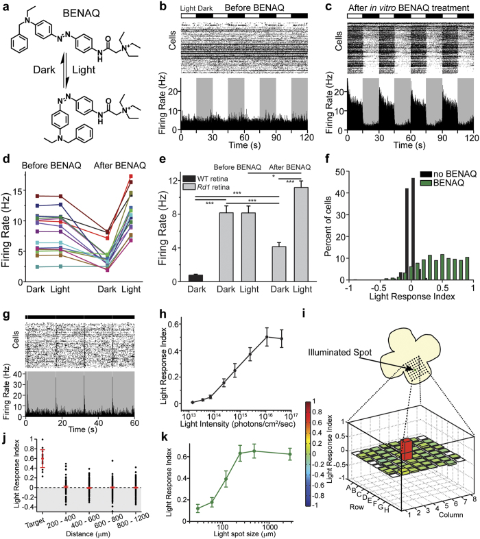
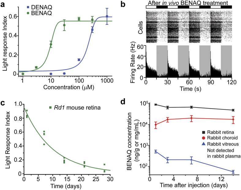
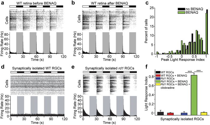
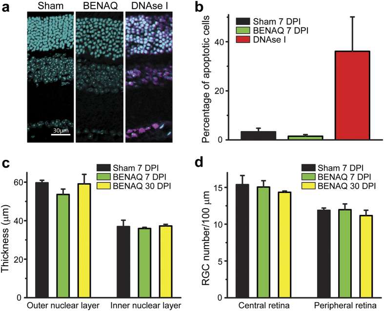
Photoswitch compounds such as DENAQ confer light-sensitivity on endogenous neuronal ion channels, enabling photocontrol of neuronal activity without genetic manipulation. DENAQ treatment restores both retinal light responses and visual behaviors in rodent models of Retinitis pigmentosa. However, retinal photosensitization requires a high dose of DENAQ and disappears within several days after treatment. Here we report that BENAQ, an improved photoswitch, is 20-fold more potent than DENAQ and persists in restoring visual responses to the retina for almost 1 month after a single intraocular injection. Studies on mice and rabbits show that BENAQ is non-toxic at concentrations 10-fold higher than required to impart light-sensitivity. These favorable properties make BENAQ a potential drug candidate for vision restoration in patients with degenerative blinding diseases.


Conflict of interest statement

R.H.K. is a member of the board of directors and J.T. is the CEO of Photoswitch Biosciences, Inc., which is developing commercial applications for chemical photoswitches. R.H.K. is an inventor on US Patent #9334231 related to using chemical photoswitches to treat blindness.

Figures

Figure 1
Figure 1. BENAQ is a photoswitch that restores spatially precise light responses to the blind retina.
(a) Structure of BENAQ. Visible light converts BENAQ from the trans to the cis form and then the compound quickly relaxes back to trans in the dark. (b,c) MEA recordings from an rd1 mouse retina before (b) and after (c) BENAQ treatment. Raster plots of individual RGC activity and average firing rate plots are shown. Alternating light (white) and dark (black) intervals plotted at the top. (d,e) Average rd1 retinal firing rate in the dark and light before (left) and after (right) BENAQ treatment (n = 16 retinas). Average WT retinal firing rate in the dark (e, left) (n = 8 retinas). Data are mean ± SEM. (f) LRI value distributions for RGCs from untreated (black) (median LRI = 0.00) and BENAQ-treated (green) rd1 retinas (median LRI = 0.51, p < 0.001, rank sum test). (g) MEA recording of a BENAQ treated rd1 mouse retina stimulated with 100 ms white light flashes every 15 seconds. (h) White light intensity – response curve for BENAQ treated rd1 retinas (n = 5 retinas). Light intensity threshold for driving RGC activity = 7 × 1013 photons/cm2/sec. Data are mean ± SEM, n = 5 retinas. (i) Rd1 retinal light response to targeted illumination of electrode E4 with a 120 μm-diameter light spot. Only electrode E4 (red) recorded an increase in RGC activity in response to white light (bottom). LRI values are color-coded (scale at left) and also represented by bar height. (j) Targeted illumination elicits an increase in activity in stimulated RGCs and has no effect on surrounding RGCs (n = 17 cells and n = 903 cells, respectively, from seven retinas). LRI values of RGCs (black circles) as a function of distance from the target electrode, displayed in 200 μm bins. Median plus and minus the 95% confidence intervals are shown in red. See also Supplementary Table S1. (k) Responses of BENAQ-treated rd1 RGCs to stimulation with light spots of increasing diameter. The light response saturates at 240 μm-diameter spot size. Data are mean ± SEM; n = 20 cells.
Figure 2
Figure 2. BENAQ is a potent, long-lasting photoswitch.
(a) Retinal photosensitization dose response curves for BENAQ (green) and DENAQ (blue). BENAQ EC50 = 9.5 μM, DENAQ EC50 = 177 μM. Data are mean ± SEM, n = 5 retinas per dose. (b) MEA recording from an rd1 mouse retina 7 days after intravitreal injection of BENAQ. (c) Time course of rd1 mouse retinal photosensitization measured via ex vivo MEA recordings at various time points after a single in vivo intravitreal injection of BENAQ. Retinal photosensitization half-life = 7.0 days, n = 4 retinas per time point. (d) Pharmacokinetic characterization of BENAQ in the rabbit eye after a single intravitreal injection of 30 μM BENAQ. BENAQ retina half-life = 24 days, BENAQ vitreous half-life = 5.4 days (extrapolated based on data up to 14 days). Data are mean ± SEM, n = 6 samples from 3 animals per time point.
Figure 3
Figure 3. BENAQ selectively photosensitizes RGCs from degenerated but not healthy retina.
(a,b) MEA recording from a WT mouse retina before (a) and after ex vivo (b) treatment with 300 μM BENAQ. (c) RGC PLRI values for WT retinas before (black) and after (green) treatment with BENAQ (n = 6 retinas, p = 0.34). (d,e) MEA recordings from BENAQ-treated pharmacologically isolated WT (d) and rd1 (e) RGCs. (f) LRI values for synaptically isolated untreated (black, n = 5 retinas) and BENAQ-treated (red, n = 5 retinas) WT RGCs, as well as for untreated (blue, n = 5 retinas), BENAQ-treated (green, n = 11 retinas) and BENAQ+ cilobradine treated (yellow, n = 4 retinas) rd1 RGCs. Data are mean ± SEM.
Figure 4
Figure 4. BENAQ has no apparent toxic effects on the mouse retina.
(a) Mouse retinal cross sections 7 Days Post Injection (DPI) of sham (left), 100 μM BENAQ (center) or after DNAse I treatment (right, positive control). Cell nuclei (cyan, DAPI) and apoptotic cells (purple, TUNEL assay) are shown. (b) Percent apoptotic cells after sham (black) or BENAQ (green) injection at 7 DPI, with DNAse I treated retinas (red) as a positive control. Data are mean ± SEM. (c) Outer nuclear layer (ONL) and inner nuclear layer (INL) thickness 7 DPI of sham (black) or BENAQ (green) and 30 DPI of BENAQ (yellow). Data are mean ± SEM. (d) RGC count per 100 μm of retina 10 DPI of sham (black) and BENAQ (green) and 30 DPI of BENAQ (yellow). Data are mean ± SEM.

References

    1. Yue L., Weiland J. D., Roska B. & Humayun M. S. Retinal stimulation strategies to restore vision: Fundamentals and systems. Progress in retinal and eye research 53, 21–47, doi: 10.1016/j.preteyeres.2016.05.002 (2016). - DOI - PubMed
    1. Jayakody S. A., Gonzalez-Cordero A., Ali R. R. & Pearson R. A. Cellular strategies for retinal repair by photoreceptor replacement. Progress in retinal and eye research 46, 31–66, doi: 10.1016/j.preteyeres.2015.01.003 (2015). - DOI - PubMed
    1. Thyagarajan S. et al.. Visual function in mice with photoreceptor degeneration and transgenic expression of channelrhodopsin 2 in ganglion cells. The Journal of neuroscience: the official journal of the Society for Neuroscience 30, 8745–8758, doi: 10.1523/jneurosci.4417-09.2010 (2010). - DOI - PMC - PubMed
    1. Busskamp V. et al.. Genetic reactivation of cone photoreceptors restores visual responses in retinitis pigmentosa. Science 329, 413–417, doi: 10.1126/science.1190897 (2010). - DOI - PubMed
    1. Polosukhina A. et al.. Photochemical restoration of visual responses in blind mice. Neuron 75, 271–282, doi: 10.1016/j.neuron.2012.05.022 (2012). - DOI - PMC - PubMed

Publication types

MeSH terms