. 2017 Jul;35(7):1860-1865.
doi: 10.1002/stem.2628. Epub 2017 Apr 24.

Brief Report: Robo1 Regulates the Migration of Human Subventricular Zone Neural Progenitor Cells During Development

Affiliations

Brief Report: Robo1 Regulates the Migration of Human Subventricular Zone Neural Progenitor Cells During Development

Hugo Guerrero-Cazares et al. Stem Cells. 2017 Jul.

Abstract

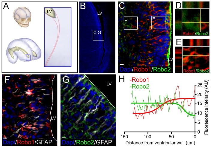
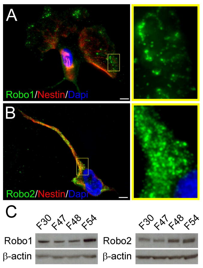
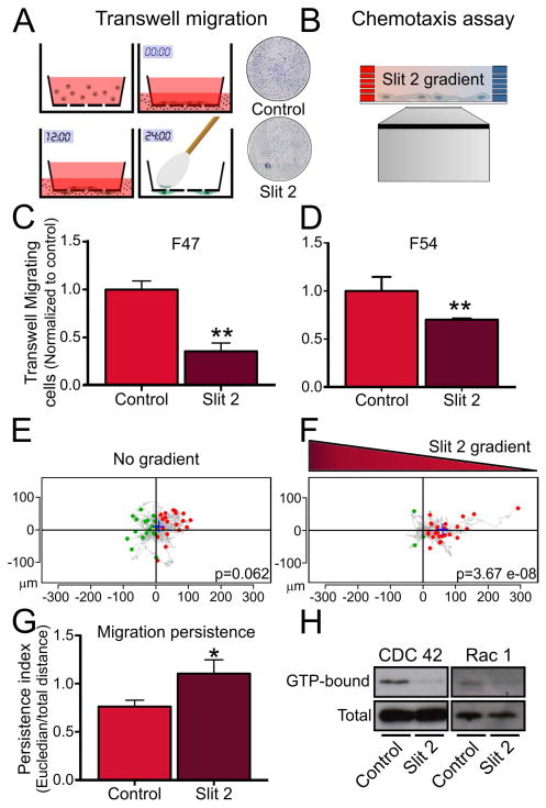
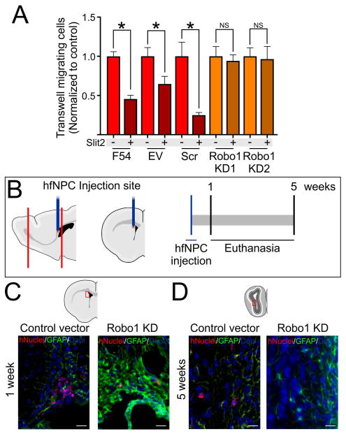
Human neural progenitor cell (NPC) migration within the subventricular zone (SVZ) of the lateral ganglionic eminence is an active process throughout early brain development. The migration of human NPCs from the SVZ to the olfactory bulb during fetal stages resembles what occurs in adult rodents. As the human brain develops during infancy, this migratory stream is drastically reduced in cell number and becomes barely evident in adults. The mechanisms regulating human NPC migration are unknown. The Slit-Robo signaling pathway has been defined as a chemorepulsive cue involved in axon guidance and neuroblast migration in rodents. Slit and Robo proteins expressed in the rodent brain help guide neuroblast migration from the SVZ through the rostral migratory stream to the olfactory bulb. Here, we present the first study on the role that Slit and Robo proteins play in human-derived fetal neural progenitor cell migration (hfNPC). We describe that Robo1 and Robo2 isoforms are expressed in the human fetal SVZ. Furthermore, we demonstrate that Slit2 is able to induce a chemorepellent effect on the migration of hfNPCs derived from the human fetal SVZ. In addition, when Robo1 expression is inhibited, hfNPCs are unable to migrate to the olfactory bulb of mice when injected in the anterior SVZ. Our findings indicate that the migration of human NPCs from the SVZ is partially regulated by the Slit-Robo axis. This pathway could be regulated to direct the migration of NPCs in human endogenous neural cell therapy. Stem Cells 2017;35:1860-1865.

Keywords: Cell migration; Human subventricular zone; Neural stem cells; Slit2.


Figures

Figure 1
Figure 1
Expression of Robo1 and Robo2 in human fetal SVZ at the LGE. A, Schematic representation of the area studied at the anterior horn of the lateral ventricle in the human fetal brain. B, Dapi stained image showing the anterior extension of the lateral ventricle and the area observed in the following frames. C-E, Co-staining of Robo1 and Robo2 isoforms shows that while Robo1 appears closer to the ventricular wall, Robo2 appears deeper into the parenchyma. F, Robo1 co-staining with GFAP shows expression of Robo1 in areas close to the ventricle where GFAP cells are present. G, Robo2 co-staining with GFAP shows a higher concentration of Robo1 deeper in the parenchyma. H, Fluorescence intensity analysis of Robo1 (red) and Robo2 (green) signals. Graph shows a higher intensity of Robo1 in areas close to the ventricle, which decreases as the distance to the ventricular wall increases. Robo2 signal intensity shows an opposite distribution, showing higher intensity in areas farther from the ventricular wall. Scale bar: 10μm
Figure 2
Figure 2
Human fetal neural progenitor cells (hfNPCs) express Robo1 and Robo2 isoforms in vitro. A ubiquitous distribution of both isoforms was observed. A-Robo1 was observed at the distal cellular projections of Nestin, expressing cells. B, Robo2 had broader distribution in the cell body but was also observed in all evaluated cell types. C, Multiple primary cultures of hfNPCs were positive for Robo1 and Robo2 isoforms by western blot analysis. Dapi was used to counterstain cell nuclei. Scale bar: 5μm.
Figure 3
Figure 3
Effects of Slit2 stimulation on hfNPCs migration. A and B, Schematic representation of a transwell migration and chemotaxis assay, respectively. C–D, Quantification of transwell migrated cells shows a reduced number of cells when Slit2 (200ng/ml) was present in the bottom well using two different primary cell cultures. E, Basal cell migration in a 2D chemotaxis assay shows aleatory migration distribution. F, hfNPCs show a biased cell migration pattern in the presence of a Slit2 gradient, indicating a chemorepellent response. G, hfNPC migration persistence is increased in response to a gradient of Slit2. H, The active forms of the small Rho-GTPases CDC42 and Rac1 are decreased upon Slit2 stimulation. **p<0.01, *p<0.05.
Figure 4
Figure 4
Effects of Robo1-KD on the migration of hfNPCs. A, Robo1-KD hfNPCs do not respond to Slit2 in a transwell migration assay. C, Schematic representation of in vivo experiment where control vector or Robo1-KD hfNPCs were injected in the anterior SVZ of 4 week old mice. D, hfNPCs at the SVZ of injected mice 1 week post injection. D, hfNPCs at the OB 5 weeks post injection. No Robo1-KD hfNPCs were observed in the OB after this term. *p<0.05 Scale bar: 20μm

References

    1. Kriegstein A, Alvarez-Buylla A. The glial nature of embryonic and adult neural stem cells. Annu Rev Neurosci. 2009;32:149–84. - PMC - PubMed
    1. Lois C, Garcia-Verdugo JM, Alvarez-Buylla A. Chain migration of neuronal precursors. Science. 1996;271(5251):978–81. - PubMed
    1. Curtis MA, et al. Human neuroblasts migrate to the olfactory bulb via a lateral ventricular extension. Science. 2007;315(5816):1243–9. - PubMed
    1. Guerrero-Cazares H, et al. Cytoarchitecture of the lateral ganglionic eminence and rostral extension of the lateral ventricle in the human fetal brain. J Comp Neurol. 2011;519(6):1165–80. - PMC - PubMed
    1. Quinones-Hinojosa A, et al. Cellular composition and cytoarchitecture of the adult human subventricular zone: a niche of neural stem cells. J Comp Neurol. 2006;494(3):415–34. - PubMed

MeSH terms

Substances