. 2017 Jul 1;158(7):2309-2318.
doi: 10.1210/en.2017-00146.

IGF1R Expression in Ovarian Granulosa Cells Is Essential for Steroidogenesis, Follicle Survival, and Fertility in Female Mice

Affiliations

IGF1R Expression in Ovarian Granulosa Cells Is Essential for Steroidogenesis, Follicle Survival, and Fertility in Female Mice

Sarah C Baumgarten et al. Endocrinology. .

Abstract

Folliculogenesis is a lengthy process that requires the proliferation and differentiation of granulosa cells (GCs) for preovulatory follicle formation. The most crucial endocrine factor involved in this process is follicle-stimulating hormone (FSH). Interestingly, previous in vitro studies indicated that FSH does not stimulate GC proliferation in the absence of the insulinlike growth factor 1 receptor (IGF1R). To determine the role of the IGF1R in vivo, female mice with a conditional knockdown of the IGF1R in the GCs were produced and had undetectable levels of IGF1R mRNA and protein in the GCs. These animals were sterile, and their ovaries were smaller than those of control animals and contained no antral follicles even after gonadotropin stimulation. The lack of antral follicles correlated with a 90% decrease in serum estradiol levels. In addition, under a superovulation protocol no oocytes were found in the oviducts of these animals. Accordingly, the GCs of the mutant females expressed significantly lower levels of preovulatory markers including aromatase, luteinizing hormone receptor, and inhibin α. In contrast, no alterations in FSH receptor expression were observed in GCs lacking IGF1R. Immunohistochemistry studies demonstrated that ovaries lacking IGF1R had higher levels of apoptosis in follicles from the primary to the large secondary stages. Finally, molecular studies determined that protein kinase B activation was significantly impaired in mutant females when compared with controls. These in vivo findings demonstrate that IGF1R has a crucial role in GC function and, consequently, in female fertility.


Figures

Figure 1.
Figure 1.
Knockdown of IGF1R expression in GCs. (a) IGF1R mRNA expression levels in GCs from eCG-treated animals expressed relative to mouse ribosomal L19 (the average of at least six samples per genotype is shown). Bars represent mean ± SEM (*P < 0.05). (b) Immunohistochemistry for the IGF1R protein in ovaries of 21- to 25-day-old controls and IGF1Rgcko eCG-stimulated mice (n = 3 for each genotype; a representative image is shown).
Figure 2.
Figure 2.
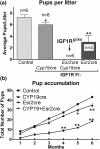
IGF1R expression in GCs is essential for female fertility. (a) The number of pups per litter was determined in control mice and experimental animals over 6 months. Four or more females were used for each genotype. Columns represent the average ± SEM of the number of pups per litter (*P < 0.05; **P < 0.01 vs. controls). IGF1Rgcko females produced no pups. (b) Continuous breeding assessment is showing the cumulative number of progeny per female. Values represent the mean ± SEM of litters derived from at least five females for each genotype (*P < 0.05; **P < 0.01 vs. controls).
Figure 3.
Figure 3.
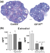
Abnormal folliculogenesis in IGF1Rgcko animals. (a) Representative hematoxylin and eosin staining of ovaries of control and IGF1Rgcko mice treated with eCG for 48 hours. (b) Estradiol levels in control (left) and experimental (right) mice treated with eCG for 48 hours. Bars represent the mean ± SEM of measurements from at least five animals per genotype. Different letters denote differences between genotypes (a–b, b–cP < 0.05; a–cP < 0.01).
Figure 4.
Figure 4.
Lack of IGF1R expression in GCs blocks ovulation. Ovulation was induced in immature (22- to 23-day-old) animals by a subcutaneous injection of 5 IU eCG, followed 48 hours later by administration of 5 IU hCG. Oviducts and ovaries were harvested 17 hours after hCG; oocytes found in the oviduct at this time were counted. Bars represent mean ± SEM for five to seven animals per group. Data were compared via t test. No oocytes were found in the oviducts of IGF1Rgcko animals.
Figure 5.
Figure 5.
Relative expression of the main differentiation markers in GCs of control and IGF1Rgcko mice. (a) Total RNA was extracted from GCs isolated after eCG treatment in control and IGF1Rgcko females. The expression of genes important for GC differentiation was measured by quantitative PCR. Three or more animals were included for each genotype. Columns represent the mean ± SEM (***P < 0.01 vs. control). (b) FSH receptor (FSHR) mRNA (left) and protein levels (right). Blots are representative of three different animals. BACT, β-actin; StAR, steroidogenic acute regulatory protein.
Figure 6.
Figure 6.
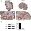
Lack of IGF1R in GCs leads to increased apoptosis and diminished Akt activation. (a) Coimmunostaining for markers of proliferation (Ki67) and apoptosis (cleaved caspase 3) were performed in control and IGF1Rgcko ovaries from 23-day-old eCG-treated mice. Cleaved caspase 3 staining is depicted in pink, Ki67 staining is depicted in brown, and counterstaining by hematoxylin is depicted in light blue. Arrowheads indicate secondary follicles, black arrows indicate early antral follicles, and yellow arrows indicate follicles with structural distortion. High magnification (×20) sections are indicated (n = 3 for each genotype; a representative picture is shown). (b) Western blots for phospho-S473 and total Akt. *P < 0.001 vs. control, n = 3.

Comment in

  • Female Fertility: It Takes Two to Tango.
    Chen LX, Jimenez PT. Chen LX, et al. Endocrinology. 2017 Jul 1;158(7):2074-2076. doi: 10.1210/en.2017-00447. Endocrinology. 2017. PMID: 28881869 Free PMC article. No abstract available.

References

    1. Chandra A, Martinez GM, Mosher WD, Abma JC, Jones J. Fertility, family planning, and reproductive health of U.S. women: data from the 2002 National Survey of Family Growth. Vital and Health Statistics Series 23. Hyattsville, MD: Centers for Disease Control and Prevention, National Center for Health Statistics; 2005:1–160. - PubMed
    1. Li RH, Ng EH. Management of anovulatory infertility. Best Pract Res Clin Obstet Gynaecol. 2012;26(6):757–768. - PubMed
    1. Kumar TR, Wang Y, Lu N, Matzuk MM. Follicle stimulating hormone is required for ovarian follicle maturation but not male fertility. Nat Genet. 1997;15(2):201–204. - PubMed
    1. Zhou P, Baumgarten SC, Wu Y, Bennett J, Winston N, Hirshfeld-Cytron J, Stocco C. IGF-I signaling is essential for FSH stimulation of AKT and steroidogenic genes in granulosa cells. Mol Endocrinol. 2013;27(3):511–523. - PMC - PubMed
    1. Baumgarten SC, Convissar SM, Fierro MA, Winston NJ, Scoccia B, Stocco C. IGF1R signaling is necessary for FSH-induced activation of AKT and differentiation of human cumulus granulosa cells. J Clin Endocrinol Metab. 2014;99(8):2995–3004. - PMC - PubMed

Publication types