. 2017 May 8;56(20):5480-5484.
doi: 10.1002/anie.201612504. Epub 2017 Apr 13.

Designed Spiroketal Protein Modulation

Affiliations

Designed Spiroketal Protein Modulation

Marcel Scheepstra et al. Angew Chem Int Ed Engl. .

Abstract

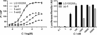
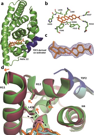
Spiroketals are structural motifs found in many biologically active natural products, which has stimulated considerable efforts toward their synthesis and interest in their use as drug lead compounds. Despite this, the use of spiroketals, and especially bisbenzanulated spiroketals, in a structure-based drug discovery setting has not been convincingly demonstrated. Herein, we report the rational design of a bisbenzannulated spiroketal that potently binds to the retinoid X receptor (RXR) thereby inducing partial co-activator recruitment. We solved the crystal structure of the spiroketal-hRXRα-TIF2 ternary complex, and identified a canonical allosteric mechanism as a possible explanation for the partial agonist behavior of our spiroketal. Our co-crystal structure, the first of a designed spiroketal-protein complex, suggests that spiroketals can be designed to selectively target other nuclear receptor subtypes.

Keywords: drug design; drug discovery; natural products; spiro compounds; structure elucidation.


Figures

Figure 1
Figure 1
Design of a spiroketal as RXR ligand. a) Molecular structures of γ‐rubromycin, [6,6]‐bisbenzannulated spiroketal 1 and RXR full agonists, BMS 649 16 and LG100268. b) Top‐ranked pose of R1 generated by docking into the space occupied by BMS 649 in the hRXRα‐BMS 649 co‐crystal structure (PDB code: 1MVC)16 using the FlexX docking module in the LeadIT suite17 followed by HYDE scoring in SEESAR.18 R1 skeleton: C=pink, O=red; protein backbone: C=green, N=blue, O=red, S=yellow; dashed lines=H‐bonding interactions below 3.3 Å.
Scheme 1
Scheme 1
Reagents and conditions: a) n‐BuLi, THF, −78 °C to RT; b) 10 % Pd/C, KHCO3, EtOAc, RT; c) Dess–Martin periodinane, CH2Cl2, RT; d) TMSBr, CH2Cl2, −30 °C to RT; e) NaOH, dioxane/MeOH, 40 °C. Characteristic 1H and 13C resonance peaks are summarized for 7 and (±)‐1.23
Figure 2
Figure 2
Biochemical and cellular evaluation of (±)‐1. Left: Fluorescence polarization assay data showing that full agonist LG100268 induces binding of the fluorescently labelled D22 peptide in a concentration‐dependent manner, while (±)‐1, 1‐ent1, and 1‐ent2 (separable by chiral HPLC) each exhibit a partial agonist behavior. Right: Cellular activities of LG100268 and (±)‐1 measured in a mammalian two‐hybrid luciferase assay. Error bars denote s.d. (n=3).
Figure 3
Figure 3
X‐ray co‐crystallography data. a) Ribbon representation of the X‐ray co‐crystal structure of R1 (orange stick) bound to the ligand‐binding pocket (LBP) of hRXRα (green ribbon) with the TIF2 co‐activator‐derived peptide (blue ribbon) PDB code: 5LYQ. b) Enlarged view of the hRXRα LBP with the amino acid side chains labelled and represented as sticks. c) Superimposition of R1 and final 2F oF c electron density map (contoured at 1σ). d) Superimposition of the LBP region of hRXRα bound to R1 (protein in green, ligand in orange, PDB code: 5LYQ) and a documented full agonist (protein in red, ligand in cyan, PDB code: 40C7).28 The TIF2 peptide corresponding to PDB code 40C7 is shown in cyan. Participating helices/residues/atoms are labelled. Orange arrows indicate movement in protein conformation from an agonistic (red) to the folded state induced by R1 (green). The loop region connecting helix 11 (H11) to H12‐443GDTPID448 is hidden for clarity.

Similar articles

References

    1. Zheng Y., Tice C. M., Singh S. B., Bioorg. Med. Chem. Lett. 2014, 24, 3673–3682. - PubMed
    1. Perron F., Albizati K. F., Chem. Rev. 1989, 89, 1617–1661.
    1. Sperry J., Wilson Z. E., Rathwell D. C. K., Brimble M. A., Nat. Prod. Rep. 2010, 27, 1117–1137. - PubMed
    1. Yoneda N., Fukata Y., Asano K., Matsubara S., Angew. Chem. Int. Ed. 2015, 54, 15497–15500; - PubMed
    2. Angew. Chem. 2015, 127, 15717–15720.
    1. Butler B. B., Manda J. N., Aponick A., Org. Lett. 2015, 17, 1902–1905. - PubMed

Publication types

LinkOut - more resources