Review
. 2017 Apr 12;21(4):433-442.
doi: 10.1016/j.chom.2017.03.010.

The Gut Microbiome: Connecting Spatial Organization to Function

Affiliations
Review

The Gut Microbiome: Connecting Spatial Organization to Function

Carolina Tropini et al. Cell Host Microbe. .

Abstract

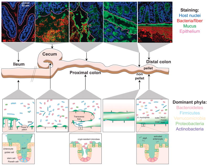
The first rudimentary evidence that the human body harbors a microbiota hinted at the complexity of host-associated microbial ecosystems. Now, almost 400 years later, a renaissance in the study of microbiota spatial organization, driven by coincident revolutions in imaging and sequencing technologies, is revealing functional relationships between biogeography and health, particularly in the vertebrate gut. In this Review, we present our current understanding of principles governing the localization of intestinal bacteria, and spatial relationships between bacteria and their hosts. We further discuss important emerging directions that will enable progressing from the inherently descriptive nature of localization and -omics technologies to provide functional, quantitative, and mechanistic insight into this complex ecosystem.

Keywords: biogeography; colon; host-microbe interactions; imaging; microbiome profiling; microbiota; mucosal.


Conflict of interest statement

Competing Financial Interests

The authors declare no competing financial interests.

Figures

Figure 1
Figure 1. Biogeography of the mouse gastrointestinal microbiota
Top: confocal micrographs of intestinal sections stained with UEA-1 (green) and DAPI (blue in epithelium, red in lumen). The UEA-1 glycan epitope is most abundant in the mouse distal colon and less so in the cecum and proximal colon. The epithelial boundary is overlaid (maroon). Middle: schematic of the distal mouse gastrointestinal tract. Bottom: schematic of key characteristics of each intestinal location after fixation with dry Carnoy’s fixative. Mucus structure and bacterial localization are heterogeneous along the longitudinal and transverse axes of the murine gastrointestinal tract. The MUC2-dependent layer becomes increasingly dense and impenetrable along the length of the intestines; the non-continuous appearance of the mucus in the ileum and proximal colon is potentially due to artefacts during mucus preparation. The density and diversity of bacteria increase along the longitudinal axis, with the small intestine favoring facultative anaerobic, proteolytic bacteria, and the colon favoring anaerobic, saccharolytic bacteria. Along the transverse axis, most bacteria are spatially segregated from the host tissue by immunological and physical barriers (Johansson et al., 2008; Vaishnava et al., 2011), with a few notable exceptions (Ivanov et al., 2009; Lee et al., 2013; Sonnenberg et al., 2012). Mucus structure in live animals is reviewed in (Pelaseyed et al., 2014). AMP, antimicrobial peptide; sIgA, secretory IgA; SFB, segmented filamentous bacteria.
Figure 2
Figure 2. Improved histological methods allow visualization of mucus heterogeneity in the mouse colon
A) Distal colon of a conventional mouse stained with UEA-1 (green), DAPI (blue in epithelium, red in lumen), and FISH probes specific to Firmicutes (yellow) and Bacteroidales (maroon). The thick, continuous, laminated inner layer of mucus adheres to the epithelium and excludes bacteria. B–C) Distal colon of a gnotobiotic mouse colonized with Bacteorides thetaiotaomicron fed a high-fiber diet (B) or polysaccharide-free diet (C) (Earle et al., 2015). The sections are stained with anti-MUC2 antibody and DAPI; images show the epithelium (blue), mucus (green), bacteria (red), and plant matter (yellow) in the lumen. The epithelial border (cyan) was identified using the software BacSpace (Earle et al., 2015). The mucus layer in (C) is thinner than in (B), likely due to its consumption by B. thetaiotaomicron.
Figure 3
Figure 3. Microscopic visualization of the gut microbiota
A) Scanning electron micrograph (Savage and Blumershine, 1974) from the mouse distal gut. Rod-, fusiform- and spiral-shaped bacteria are present, illustrating the morphological diversity of the gut microbiota. B) Scanning electron micrograph from the mouse colon (Savage and Blumershine, 1974), highlighting the high density of bacteria in the gut. C) Inter-kingdom spatial interactions between helminths and bacteria have been poorly studied to date. Trichuris muris, a mouse model of whipworm, was visualized by labeling with WGA (red) in the proximal colon of a conventional Swiss-Webster mouse 14 days after inoculation with ~200 T. muris ova. The anterior end is embedded in the epithelium near gastrointestinal lymphoid tissue (white arrowheads), while the posterior end is free in the lumen. MUC2 is labeled in green, and the mouse and worm nuclei are labeled with DAPI in blue. D) Magnification of white box in (C) with bacterial DAPI signal segmented from the worm DAPI signal and false-colored yellow. Bacteria can be seen embedded in the cuticle of the worm. E) Much of what we know about localization in the gut is derived from mouse studies. Further studies of human biopsies are needed, and in particular, on samples with preserved mucus. Here, a biopsy from a healthy patient has been fixed in methacarn, processed, sectioned and stained as described in Box 1. Bacteria, which are labeled with DAPI and false-colored red, are visible on the luminal side of the inner mucus layer.
Figure 4
Figure 4. Future directions and challenges for localization studies of the gut microbiota
Biogeography studies require technological and methodological development from the organismal/organ scale (left) to the cellular and sub-cellular scales (right). Highlighted are some of the potential challenges and improvements that will advance the field of microbiota organization.

References

    1. Alam A, Leoni G, Quiros M, Wu H, Desai C, Nishio H, Jones RM, Nusrat A, Neish AS. The microenvironment of injured murine gut elicits a local pro-restitutive microbiota. Nature Microbiology. 2016;1:15021. - PMC - PubMed
    1. Albenberg L, Esipova TV, Judge CP, Bittinger K, Chen J, Laughlin A, Grunberg S, Baldassano RN, Lewis JD, Li H, et al. Correlation between intraluminal oxygen gradient and radial partitioning of intestinal microbiota. Gastroenterology. 2014;147:1055–1063. e1058. - PMC - PubMed
    1. Amar J, Chabo C, Waget A, Klopp P, Vachoux C, Bermudez-Humaran LG, Smirnova N, Berge M, Sulpice T, Lahtinen S, et al. Intestinal mucosal adherence and translocation of commensal bacteria at the early onset of type 2 diabetes: molecular mechanisms and probiotic treatment. EMBO molecular medicine. 2011;3:559–572. - PMC - PubMed
    1. Ambort D, Johansson ME, Gustafsson JK, Nilsson HE, Ermund A, Johansson BR, Koeck PJ, Hebert H, Hansson GC. Calcium and pH-dependent packing and release of the gel-forming MUC2 mucin. Proceedings of the National Academy of Sciences of the United States of America. 2012;109:5645–5650. - PMC - PubMed
    1. Arena ET, Campbell-Valois FX, Tinevez JY, Nigro G, Sachse M, Moya-Nilges M, Nothelfer K, Marteyn B, Shorte SL, Sansonetti PJ. Bioimage analysis of Shigella infection reveals targeting of colonic crypts. Proceedings of the National Academy of Sciences of the United States of America. 2015;112:E3282–3290. - PMC - PubMed