. 2017 Apr 13;17(1):99.
doi: 10.1186/s12872-017-0530-5.

Global Bi-ventricular endocardial distribution of activation rate during long duration ventricular fibrillation in normal and heart failure canines

Affiliations

Global Bi-ventricular endocardial distribution of activation rate during long duration ventricular fibrillation in normal and heart failure canines

Qingzhi Luo et al. BMC Cardiovasc Disord. .

Abstract

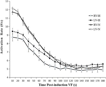
Background: The objective of this study was to detect differences in the distribution of the left and right ventricle (LV & RV) activation rate (AR) during short-duration ventricular fibrillation (SDVF, <1 min) and long-duration ventricular fibrillation VF (LDVF, >1 min) in normal and heart failure (HF) canine hearts.

Methods: Ventricular fibrillation (VF) was electrically induced in six healthy dogs (control group) and six dogs with right ventricular pacing-induced congestive HF (HF group). Two 64-electrode basket catheters deployed in the LV and RV were used for global endocardium electrical mapping. The AR of VF was estimated by fast Fourier transform analysis from each electrode.

Results: In the control group, the LV was activated faster than the RV in the first 20 s, after which there was no detectable difference in the AR between them. When analyzing the distribution of the AR within the bi-ventricles at 3 min of LDVF, the posterior LV was activated fastest, while the anterior was slowest. In the HF group, a detectable AR gradient existed between the two ventricles within 3 min of VF, with the LV activating more quickly than the RV. When analyzing the distribution of the AR within the bi-ventricles at 3 min of LDVF, the septum of the LV was activated fastest, while the anterior was activated slowest.

Conclusions: A global bi-ventricular endocardial AR gradient existed within the first 20 s of VF but disappeared in the LDVF in healthy hearts. However, the AR gradient was always observed in both SDVF and LDVF in HF hearts. The findings of this study suggest that LDVF in HF hearts can be maintained differently from normal hearts, which accordingly should lead to the development of different management strategies for LDVF resuscitation.

Keywords: Activation Rate; Heart Failure; Ventricular Fibrillation.


Figures

Fig. 1
Fig. 1
Global electrical mapping of the RV and LV endocardium. Panel I shows a fluoroscopic image of a posterior–anterior view of two basket catheters in the LV and RV and the RV defibrillation catheter. Panel II shows the basket orientation in the LV and RV. R, right free wall; A, anterior free wall; L, left free wall; P, posterior free wall; and S, septum. Apical electrodes are placed toward the center of the display, and basal electrodes are located near the periphery
Fig. 2
Fig. 2
Evolution of the AR during VF. In the control group, there was only an AR gradient between the ventricles within the first 20 s. In contrast, in the HF group, there was a detectable bi-ventricular AR gradient for the entire VF duration. From 90 s to the end of analysis at 3 min of VF, neither the RV or LV in the HF hearts activated differently than those of the control animals. (LV-N: left ventricle in the normal group; LV-H: left ventricle in the HF group; RV-N: right ventricle in the normal group; RV-H: right ventricle in the HF group.) See the text of the article for additional details
Fig. 3
Fig. 3
Snapshots of activation during VF in one normal and one HF-affected dog at the 3 min LDVF. Recordings of VF activation from one control heart are shown in Fig. 3 a and b, with Fig. 3 a I representing the posterior wall and II representing the anterior wall of the LV. In Fig. 3 b, I indicates the posterior wall, and II indicates the anterior wall of RV; as shown, the posterior wall activates faster than the anterior wall. Recordings of VF activation from one HF-affected animal are shown in Fig. 3 c and d. Figure 3 c I shows the septal wall, and II shows the anterior wall of the LV. Figure 3 d I shows the septal wall, and II indicates the anterior wall of the RV; the septal wall activates faster than the anterior wall
Fig. 4
Fig. 4
3 min LDVF regional AR distribution in the RV and LV in one normal and one HF animal. Figure 4 I shows the regional AR distribution of the two ventricles in the control group for the 3 min LDVF. In the LV, the posterior wall activates before the anterior wall. In the RV, the posterior wall also activates before the anterior wall. Figure 4 II represents the regional AR distribution of the RV and LV in the HF group for the 3 min LDVF. In the LV, the septal tissue activates the fastest, while the anterior wall activates the slowest. In the RV, the septal tissue activates the fastest, while the anterior wall activates the slowest. The colors represent the AR of the 64-basket electrodes according to the time scale shown to the right (blue represents the fastest activation and red represents the slowest activation)
Fig. 5
Fig. 5
Three-minute LDVF AR distribution of the apical and basal portions of the bi-ventricles. Figure 5 a represents the apex-base AR differences in the control group, with the apex activating faster than the base in the LV (*P<0.01) and RV (*P<0.01). Figure 5 b shows the apex-base AR differences in the HF group, with the apex activating faster than the base in the LV (*P<0.01) and RV (*P<0.01)

Similar articles

References

    1. Choi B-R, Nho W, Liu T, Salama G. Life span of ventricular fibrillation frequencies. Circ Res. 2002;91(4):339–345. doi: 10.1161/01.RES.0000031801.84308.F4. - DOI - PubMed
    1. Valderrábano M, Yang J, Omichi C, et al. Frequency analysis of ventricular fibrillation in swine ventricles. Circ Res. 2002;90(2):213–222. doi: 10.1161/hh0202.103645. - DOI - PubMed
    1. Moreno J, Zaitsev AV, Warren M, et al. Effect of remodelling, stretch and ischaemia on ventricular fibrillation frequency and dynamics in a heart failure model. Cardiovasc Res. 2005;65(1):158–166. doi: 10.1016/j.cardiores.2004.09.006. - DOI - PubMed
    1. Nanthakumar K, Huang J, Rogers JM, et al. Regional differences in ventricular fibrillation in the open-chest porcine left ventricle. Circ Res. 2002;91(8):733–740. doi: 10.1161/01.RES.0000038945.66661.21. - DOI - PubMed
    1. Umapathy K, Masse S, Sevaptsidis E, et al. Regional frequency variation during human ventricular fibrillation. Med Eng Phys. 2009;31(8):964–970. doi: 10.1016/j.medengphy.2009.05.009. - DOI - PubMed

Publication types

MeSH terms