. 2017 Apr 13;13(1):102.
doi: 10.1186/s12917-017-1028-1.

Amyloid precursor protein, lipofuscin accumulation and expression of autophagy markers in aged bovine brain

Affiliations

Amyloid precursor protein, lipofuscin accumulation and expression of autophagy markers in aged bovine brain

D De Biase et al. BMC Vet Res. .

Abstract

Background: Autophagy is a highly regulated process involving the bulk degradation of cytoplasmic macromolecules and organelles in mammalian cells via the lysosomal system. Dysregulation of autophagy is implicated in the pathogenesis of many neurodegenerative diseases and integrity of the autophagosomal - lysosomal network appears to be critical in the progression of aging. Our aim was to survey the expression of autophagy markers and Amyloid precursor protein (APP) in aged bovine brains. For our study, we collected samples from the brain of old (aged 11-20 years) and young (aged 1-5 years) Podolic dairy cows. Formalin-fixed and paraffin embedded sections were stained with routine and special staining techniques. Primary antibodies for APP and autophagy markers such as Beclin-1 and LC3 were used to perform immunofluorescence and Western blot analysis.

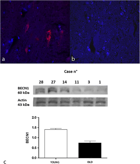
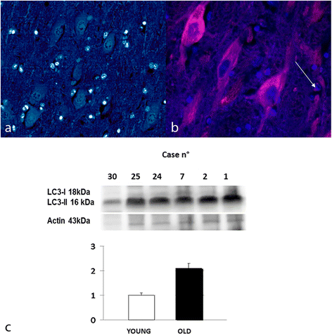
Results: Histologically, the most consistent morphological finding was the age-related accumulation of intraneuronal lipofuscin. Furthermore, in aged bovine brains, immunofluorescence detected a strongly positive immunoreaction to APP and LC3. Beclin-1 immunoreaction was weak or absent. In young controls, the immunoreaction for Beclin-1 and LC3 was mild while the immunoreaction for APP was absent. Western blot analysis confirmed an increased APP expression and LC3-II/LC3-I ratio and a decreased expression of Beclin-1 in aged cows.

Conclusions: These data suggest that, in aged bovine, autophagy is significantly impaired if compared to young animals and they confirm that intraneuronal APP deposition increases with age.

Keywords: Ageing; Autophagy; Bovine; Brain; Neuropathology.


Figures

Fig. 1
Fig. 1
a Lipofuscin storage, hippocampus, dentate gyrus. Pyramidal neurons showing an abundant granular, PAS positive, intracytoplasmatic storage material. Periodic acid-Schiff (PAS) stain, 40X. b Intraneuronal storage material exhibits green-yellow autofluorescence by fluorescence microscopy. Fluorescence microscope; FITC filter (excitation, 455–500 nm; emission, 500–570 nm), 40X. c Neurons of young controls does not exhibit PAS positive and intracytoplasmatic storage material. Periodic acid-Schiff (PAS) stain, 40X
Fig. 2
Fig. 2
Beclin 1 expression, hippocampus, dentate gyrus. a Immunostaining inyoung and b aged brain. TRITC filter (excitation, 543 nm; emission 560 nm). DAPI counterstain, 40X. c Protein expression in young and aged bovine brains. Densitometric values shows that BECN1 is expressed in higher amount in young controls when compared to aged brain samples (P < 0,001 vs controls). Bars refers to mean values. Actin protein levels confirm the amount of protein loading in each lane
Fig. 3
Fig. 3
LC3 expression, hippocampus, dentate gyrus. a Immunostaining in young and b aged brain. Immunoreaction for LC3 was detected also in astrocytes (arrow).TRITC filter (excitation, 543 nm; emission 560 nm). DAPI counterstain, 40X. c Protein expression in young and aged bovine brains. Densitometric values shows that LC3-II/LC3-I ratio is significantly increased in the aged animals compared to the young controls (P < 0,05 vs control). Bars refers to mean values. Actin protein levels confirm the amount of protein loading in each lane
Fig. 4
Fig. 4
APP expression, hippocampus, dentate gyrus. a Immunostaining in young and b aged brain. TRITC filter (excitation, 543 nm; emission 560 nm). DAPI counterstain, 40X. c Protein expression in young and aged bovine brains. Densitometric values shows that APP is expressed in higher amount in aged brains when compared to young controls (P < 0,05 vs controls). Bars refers to mean values. Actin protein levels confirm the amount of protein loading in each lane

Similar articles

Cited by

References

    1. Wojcik S. Crosstalk between autophagy and proteasome protein degradation systems: possible implications for cancer therapy. Folia Histochem Cytobiol. 2013;51(4):249–264. doi: 10.5603/FHC.2013.0036. - DOI - PubMed
    1. Mizushima N. Autophagy: Process and function. Genes Dev. 2007;21:2861–2873. doi: 10.1101/gad.1599207. - DOI - PubMed
    1. Kim PK, Hailey DW, Mullen RT, Lippincott-Schwartza J. Ubiquitin signals autophagic degradation of cytosolic proteins and peroxisomes. Proc Natl Acad Sci U S A. 2008;105(52):20567–20574. doi: 10.1073/pnas.0810611105. - DOI - PMC - PubMed
    1. Cuervo AM, Bergamini E, Brunk UT, et al. Autophagy and aging: the importance of maintaining “clean” cells. Autophagy. 2005;1:131–140. doi: 10.4161/auto.1.3.2017. - DOI - PubMed
    1. Selkoe DJ, Podlisny MB, Joachim CL, Vickers EA, Lee G, Fritz LC, Oltersdorf T. Beta-amyloid precursor protein of Alzheimer disease occurs as 110- to 135-kilodalton membrane-associated proteins in neural and non neural tissues. Proc Nat Acad Sci U S A. 1988;85(19):7341–7345. doi: 10.1073/pnas.85.19.7341. - DOI - PMC - PubMed