. 2017 Apr 13;36(1):53.
doi: 10.1186/s13046-017-0528-y.

Exosomal transfer of tumor-associated macrophage-derived miR-21 confers cisplatin resistance in gastric cancer cells

Affiliations

Exosomal transfer of tumor-associated macrophage-derived miR-21 confers cisplatin resistance in gastric cancer cells

Peiming Zheng et al. J Exp Clin Cancer Res. .

Abstract

Background: Cisplatin-based chemotherapy is frequently used to treat advanced gastric cancer (GC). However, the resistance often occurs with the mechanisms being not well understood. Recently, emerging evidence indicates that tumor-associated macrophages (TAMs) play an important role in chemoresistance of cancer. As the important mediators in intercellular communications, exosomes secreted by host cells mediate the exchange of genetic materials and proteins to be involved in tumor aggressiveness. The aim of the study was to investigate whether exosomes derived from TAMs mediate cisplatin resistance in gastric cancer.

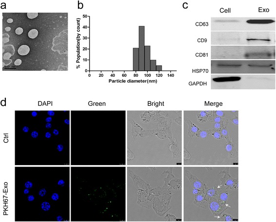
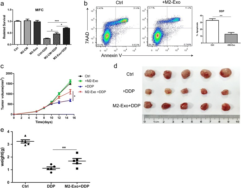
Methods: M2 polarized macrophages were obtained from mouse bone marrow or human PBMCs stimulated with IL-4 and IL-13. Exosomes isolated from M2 macrophages culture medium were characterized, and miRNA expression profiles of M2 derived exosomes (M2-exos) were analyzed using miRNA microarray. In vitro cell coculture was further conducted to investigate M2-exos mediated crosstalk between TAMs and tumor cells. Moreover, the in vivo experiments were performed using a subcutaneous transplantation tumor model in athymic nude mice.

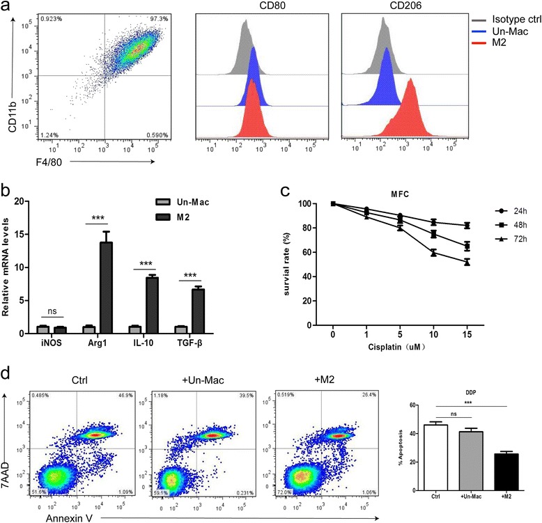
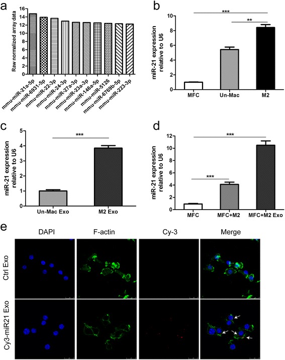
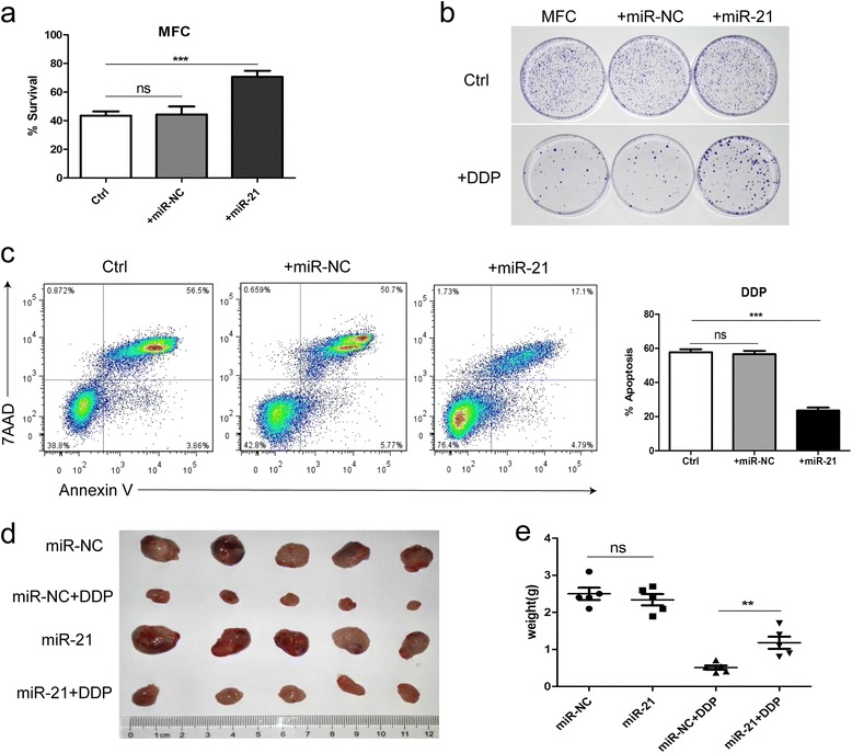
Results: In this study, we showed that M2 polarized macrophages promoted cisplatin (DDP) resistance in gastric cancer cells and exosomes derived from M2 macrophages (M2-exos) are involved in mediating the resistance to DDP. Using miRNA profiles assay, we identify significantly higher levels of microRNA-21 (miR21) isomiRNAs in exosomes and cell lysate isolated from M2 polarized macrophage. Functional studies revealed that exosomal miR-21 can be directly transferred from macrophages to the gastric cancer cells, where it suppresses cell apoptosis and enhances activation of PI3K/AKT signaling pathway by down-regulation of PTEN.

Conclusions: Our findings suggest that exosomal transfer of tumor-associated macrophages derived miR-21 confer DDP resistance in gastric cancer, and targeting exosome communication may be a promising new therapeutic strategy for gastric cancer patients.

Keywords: Cisplatin resistance; Exosome; Gastric cancer; Tumor-associated macrophages; miR-21.


Figures

Fig. 1
Fig. 1
Co-cultivation with M2 polarized macrophages enhances the resistance of MFC cells to cisplatin. a FCM identification of M2 polarized macrophages derived from murine bone marrow stimulated with IL-4 and IL-13, CD206 was a specific marker for M2. (Un-Mac, unactivated macrophages; M2, macrophages activated by IL-4 and IL-13). b qRT-PCR detection of iNOS, Arg1, IL-10, TGF-β mRNA expression in unactivated and M2 polarized macrophages, GAPDH was assayed as an internal control. (ns p > 0.05, *** p < 0.001). c Cell viability assay of MFC treated with various concentrations of DDP for 24 h, 48 h, and 72 h. d Flow cytometric analyses of apoptotic cells. MFC cells were cultured alone or co-cultured with unactivated (Un-Mac) or M2 polarized macrophages, and then exposed to DDP for 48 h. The quantitative data are presented as the mean ± SD of triplicate experiments. (ns p > 0.05, *** p < 0.001)
Fig. 2
Fig. 2
Exosomes derived from M2 polarized macrophages can be internalized by gastric cancer cells. a Representative transmission electron microscopy image of M2-derived exosomes (scale bar, 100 nm). b Histogram showing the particle diameter (nm) of the purified exosomes. c Exosomal markers (CD63, CD9, CD81, HSP70) were analyzed in M2 cellular protein and corresponding exosomes using western blotting (GAPDH was used as an internal reference). d The uptake of the PKH67 labelled M2-Exos was evident in MFCcells after 12 h of incubation. No stain was observed in the negative control condition. (scale bar, 10 um)
Fig. 3
Fig. 3
M2-exos induce the resistance of MFC cells to DDP in vitro and in vivo. a Cell viability was assessed by CCK-8 assays. M2-derived conditioned medium (CM) or exosomes (M2-Exo) attenuated DDP-induced cell suppression. MFC cells were pretreated with M2-derived CM or exosomes, and then exposed to DDP for 48 h. b Flow cytometric analyses of apoptotic cells. MFC cells were exposed to DDP alone (ctrl) or DDP and M2-Exo for 48 h. The quantitative data are presented as the mean ± SD of triplicate experiments. (*** p < 0.001). c Tumor growth curves in nude mice inoculated with MFC cells that were pretreated with or without M2-Exo.DDP or PBS (ctrl) treatment was initiated 10 days after subcutaneous injections of tumor cells. (n = 5, * p < 0.05). d The size of tumors at the end of the experiment from mice treated with PBS (ctrl), DDP, M2-Exo + DDP. (n = 5). e The mean weight of tumors from mice shown in (d). (n = 5, ** p < 0.01)
Fig. 4
Fig. 4
Exosomal transfer of miR-21 from M2 macrophages to gastric cancer cells. a The top 10 most abundant miRNAs in M2-Exo are listed, and miR-21 was the most abundant. b qRT-PCR detection of miR-21 in MFC, unactivated macrophages (Un-Mac) and M2 polarized macrophages (M2). (**p < 0.01, *** p < 0.001). c qRT-PCR detection of miR-21 in exosomes derived from unactivated macrophages (Un-Mac) and M2 polarized macrophages (M2). (*** p < 0.001). d qRT-PCR detection of miR-21 in MFC cultured alone (ctrl) or co-cultured with M2 macrophages or M2-Exo. (*** p < 0.001). e Exosomes prepared from M2 macrophages transfected with Cy3-labelled miR-21 or without transfection (ctrl) were added to MFC cell cultures. MFC cells were fixed and stained with DAPI and Alexa 488 phalloidin. The fluorescence signals in MFC cells were detected by SP5 confocal microscope. (scale bar, 25um)
Fig. 5
Fig. 5
miR-21 induces the resistance of MFC cells to DDP in vitro and in vivo. a Cell viability assay of MFC cells treated with DDP after transfected with miR-21 mimics or miR-NC or without transfection (ctrl). b Colony formation assay of MFC cells transfected with miR-21 mimics or miR-NC or without transfection (up), and exposed to DDP (below). c Cell apoptosis assay of MFC cells treated with DDP after transfected with miR-21 mimics or miR-NC or without transfection (ctrl). The quantitative data are presented as the mean ± SD of triplicate experiments. (ns p > 0.05,*** p < 0.001). d and e The size and mean weight of tumors at the end of the experiment from mice inoculated with MFC cells transfected with miR-21 mimics or miR-NC. DDP or PBS (ctrl) treatment was initiated 10 days after subcutaneous injections of tumor cells. (n = 5, ns p > 0.05, ** p < 0.01)
Fig. 6
Fig. 6
Exosomal transfer of miR-21 enhances gastric cancer cells chemoresistance via the PTEN/PI3K/AKT signaling pathway. a qRT-PCR detection of PTEN in MFC cells treated with or without M2-Exo. (*** p < 0.001). b Western blot assays for the expression of PI3Kp85, PTEN, p-AKT, AKT, BCL-2 in MFC cells treated with or without M2-Exo.GAPDH was used as the loading control. c qRT-PCR detection of PTEN in MFC cells transfected with miR-21 mimics or miR-NC or without transfection (ctrl). d Western blot assays for the expression of PI3Kp85, PTEN, p-AKT, AKT, BCL-2 in MFC cells transfected with miR-21 mimics or miR-NC or without transfection (ctrl). GAPDH was used as the loading control

References

    1. Torre LA, Bray F, Siegel RL, Ferlay J, Lortet-Tieulent J, Jemal A. Global cancer statistics, 2012. CA Cancer J Clin. 2015;65:87–108. doi: 10.3322/caac.21262. - DOI - PubMed
    1. Chen W, Zheng R, Baade PD, Zhang S, Zeng H, Bray F, Jemal A, Yu XQ, He J. Cancer statistics in China, 2015. CA Cancer J Clin. 2016;66:115–32. doi: 10.3322/caac.21338. - DOI - PubMed
    1. Orditura M, Galizia G, Sforza V, Gambardella V, Fabozzi A, Laterza MM, Andreozzi F, Ventriglia J, Savastano B, Mabilia A, et al. Treatment of gastric cancer. World J Gastroenterol. 2014;20:1635–49. doi: 10.3748/wjg.v20.i7.1635. - DOI - PMC - PubMed
    1. Gajewski TF, Schreiber H, Fu YX. Innate and adaptive immune cells in the tumor microenvironment. Nat Immunol. 2013;14:1014–22. doi: 10.1038/ni.2703. - DOI - PMC - PubMed
    1. Quail DF, Joyce JA. Microenvironmental regulation of tumor progression and metastasis. Nat Med. 2013;19:1423–37. doi: 10.1038/nm.3394. - DOI - PMC - PubMed

Publication types