. 2017 Jun 7;25(6):1387-1394.
doi: 10.1016/j.ymthe.2017.03.029. Epub 2017 Apr 10.

5 Year Expression and Neutrophil Defect Repair after Gene Therapy in Alpha-1 Antitrypsin Deficiency

Affiliations

5 Year Expression and Neutrophil Defect Repair after Gene Therapy in Alpha-1 Antitrypsin Deficiency

Christian Mueller et al. Mol Ther. .

Abstract

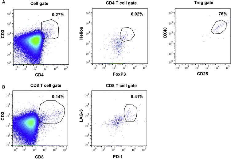
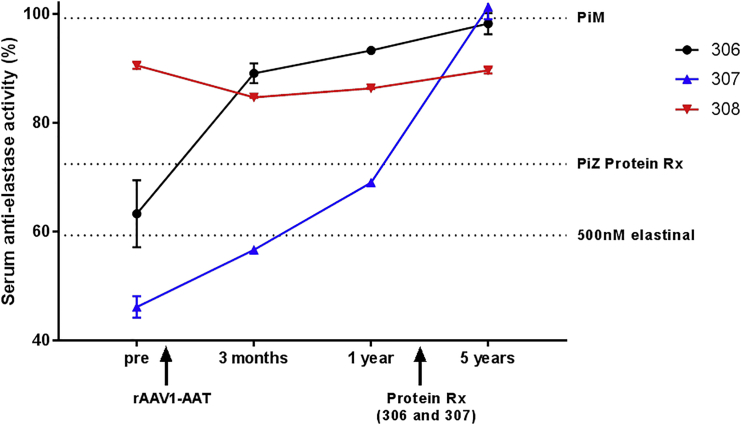
Alpha-1 antitrypsin deficiency is a monogenic disorder resulting in emphysema due principally to the unopposed effects of neutrophil elastase. We previously reported achieving plasma wild-type alpha-1 antitrypsin concentrations at 2.5%-3.8% of the purported therapeutic level at 1 year after a single intramuscular administration of recombinant adeno-associated virus serotype 1 alpha-1 antitrypsin vector in alpha-1 antitrypsin deficient patients. We analyzed blood and muscle for alpha-1 antitrypsin expression and immune cell response. We also assayed previously reported markers of neutrophil function known to be altered in alpha-1 antitrypsin deficient patients. Here, we report sustained expression at 2.0%-2.5% of the target level from years 1-5 in these same patients without any additional recombinant adeno-associated virus serotype-1 alpha-1 antitrypsin vector administration. In addition, we observed partial correction of disease-associated neutrophil defects, including neutrophil elastase inhibition, markers of degranulation, and membrane-bound anti-neutrophil antibodies. There was also evidence of an active T regulatory cell response (similar to the 1 year data) and an exhausted cytotoxic T cell response to adeno-associated virus serotype-1 capsid. These findings suggest that muscle-based alpha-1 antitrypsin gene replacement is tolerogenic and that stable levels of M-AAT may exert beneficial neutrophil effects at lower concentrations than previously anticipated.

Keywords: A1AT; AAT; AAV; PD-1; Tregs; alpha-1 antitrypsin; clinical trial; exhausted T cells; gene therapy; rAAV.


Figures

Figure 1
Figure 1
M-AAT Serum Levels over the 5 Years after Intramuscular Administration of rAAV1-CB-hAAT The dotted line is at ∼2.5% of the target therapeutic serum concentration of 572 μg/mL (11 μM). The serum AAT levels were assayed using a M-AAT-specific ELISA. The high dose was 6.0 × 1012 vg/kg, the mid dose was 1.9 × 1012 vg/kg, and the low dose was 6.0 × 1011 vg/kg. Early time points previously reported in Mueller et al.
Figure 2
Figure 2
M-AAT Expression in Skeletal Muscle 3 Months, 12 Months, and 5 Years after Intramuscular Administration of rAAV1-CB-hAAT Muscle immunochemistry staining for hAAT. The specimens were biopsied from the area injected with rAAV1 and the tissue stained for hAAT (brown). The AAT expression was persistent, while inflammatory cell infiltrates decreased over time from 3 months to 5 years. All of the slides are from patient 308. (A) Tissue 3 months after rAAV1-CB-hAAT delivery. (B) Tissue 12 months after rAAV1-CB-hAAT delivery. (C and D) Tissue 5 years after rAAV1-CB-hAAT delivery.
Figure 3
Figure 3
Immunophenotyping of Cellular Infiltrates in Skeletal Muscle at the Injection Site 5 Years after Intramuscular Administration of rAAV1-CB-hAAT (A) hAAT immunohistochemistry showing microfoci of immune cells around cell expression hAAT. (B) CD3 staining, demonstrating that many of the mononuclear immune cells are T-lymphocytes. (C) CD8 staining indicating a population of cytotoxic T cells. (D) CD68 staining indicating a population of macrophages. (E) CD20 staining indicating a small population of B-lymphocytes.
Figure 4
Figure 4
Bisulfite Sequencing to Quantify the Percentage of Immune Cells in Muscle Biopsies Obtained 5 Years after Intramuscular Administration of rAAV1-CB-hAAT A relatively high proportion of Tregs was observed in the muscle immune cell population. (A) Percentage of CD3+, Treg (FOXP3+), and CD8+ cells in the muscle. (B) Treg and CD8+ cells as a percentage of CD3+ cell population in the muscle.
Figure 5
Figure 5
Regulatory T Cell and Exhausted T Cell Detection by Flow Cytometry in Samples from Muscle Biopsies Obtained 5 Years after Intramuscular Administration of rAAV1-CB-hAAT (A) Regulatory T cells were gated as Helios+FoxP3+ cells in the CD3+CD4+ subset. The activated Treg cells were defined as OX40+CD25+ cells. (B) Exhausted CD8 T cells were gated as LAG-3+PD-1+ cells in the CD3+CD8+ subset. The percentages of each population are indicated in the dot plots.
Figure 6
Figure 6
Peripheral Gamma-IFN ELISPOT Responses to AAV Capsid Epitope Pools A decreased peripheral blood AAV1 reactivity was observed at month 52 compared to earlier time points (all of the data from patient 308). SFU, spot forming units; PBMC, peripheral blood mononuclear cell; AAV1a/b/c, pools of AAV1 peptides; CEF, positive control peptide pool.
Figure 7
Figure 7
Elastase Inhibitory Activity Assay Inhibition of neutrophil elastase was quantified by a colorimetric assay in triplicates and reported as % of no inhibitor control ±SEM. The patients were dosed with rAAV1-AAT gene therapy treatment at day 0 and serum was sampled pretreatment and at 3 months, 1 year, and 5 years post treatment. Patients 306 and 307 started protein augmentation therapy (Protein Rx) after their 1 year follow up. Additional controls included two PiM (MM) individuals and two PiZ (ZZ) individuals under protein augmentation therapy (PiZ Protein Rx) (500 nM Elastinal indicates the elastase activity control).
Figure 8
Figure 8
Biomarkers of Degranulation and Auto-Antibodies: TNF-R1 and Neutrophil Anti-Lactoferrin TNF-R1 levels in patients treated with gene therapy approached that of control PiM patients (M-AAT/MM). Anti-lactoferrin staining indicated that the rAAV1-AAT treated patient had an approximately 50% correction in this marker. (A) TNFR1 levels in the serum 5 years post-gene delivery. The patient 306 serum data were collected following lung transplant and subsequent pulmonary Aspergillosis diagnosis. (B) Anti-lactoferrin levels in PFA fixed neutrophils (AAT Day 2: Day 2 after protein replacement therapy in a PiZ [ZZ] patient).

References

    1. de Serres F., Blanco I. Role of alpha-1 antitrypsin in human health and disease. J. Intern. Med. 2014;276:311–335. - PubMed
    1. Larsson C. Natural history and life expectancy in severe alpha1-antitrypsin deficiency, Pi Z. Acta Med. Scand. 1978;204:345–351. - PubMed
    1. Browne R.J., Mannino D.M., Khoury M.J. Alpha 1-antitrypsin deficiency deaths in the United States from 1979-1991. An analysis using multiple-cause mortality data. Chest. 1996;110:78–83. - PubMed
    1. Aliño S.F., Crespo J., Bobadilla M., Lejarreta M., Blaya C., Crespo A. Expression of human alpha 1-antitrypsin in mouse after in vivo gene transfer to hepatocytes by small liposomes. Biochem. Biophys. Res. Commun. 1994;204:1023–1030. - PubMed
    1. Aliño S.F., Bobadilla M., Crespo J., Lejarreta M. Human alpha 1-antitrypsin gene transfer to in vivo mouse hepatocytes. Hum. Gene Ther. 1996;7:531–536. - PubMed

Publication types

MeSH terms

Supplementary concepts