. 2017 Aug;7(8):852-867.
doi: 10.1158/2159-8290.CD-16-1020. Epub 2017 Apr 13.

Synergistic Immunostimulatory Effects and Therapeutic Benefit of Combined Histone Deacetylase and Bromodomain Inhibition in Non-Small Cell Lung Cancer

Affiliations

Synergistic Immunostimulatory Effects and Therapeutic Benefit of Combined Histone Deacetylase and Bromodomain Inhibition in Non-Small Cell Lung Cancer

Dennis O Adeegbe et al. Cancer Discov. 2017 Aug.

Abstract

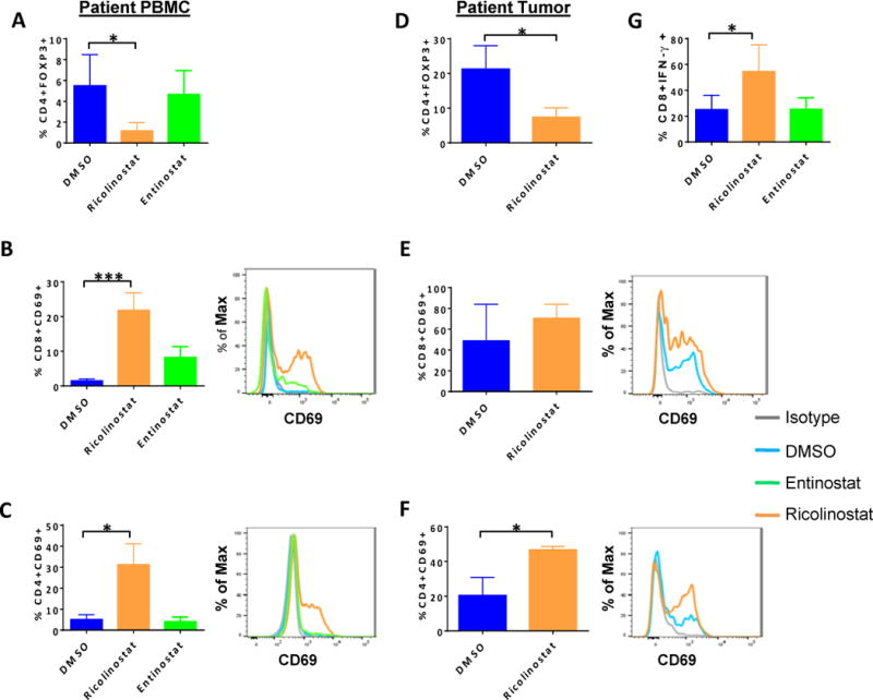
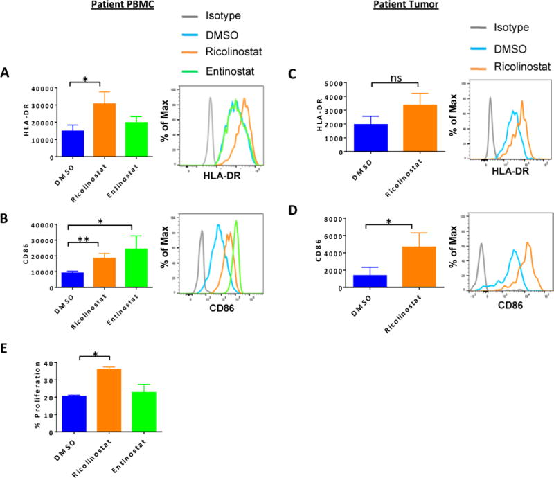
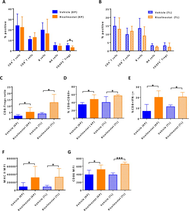
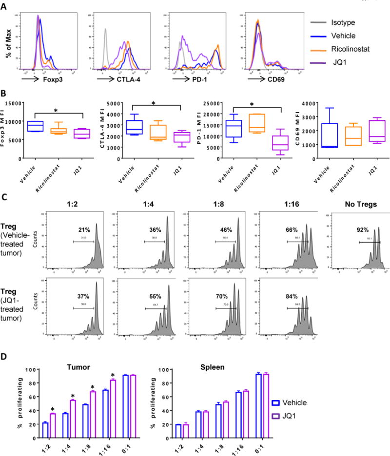
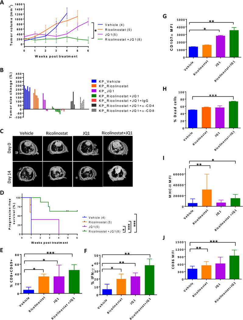
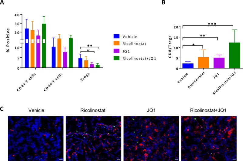
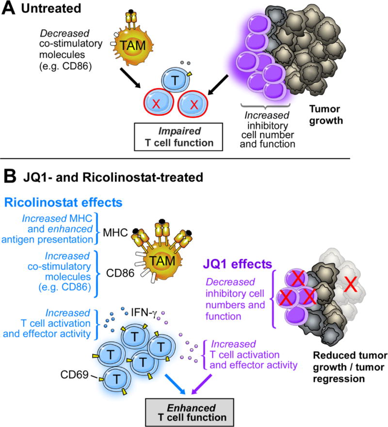
Effective therapies for non-small cell lung cancer (NSCLC) remain challenging despite an increasingly comprehensive understanding of somatically altered oncogenic pathways. It is now clear that therapeutic agents with potential to impact the tumor immune microenvironment potentiate immune-orchestrated therapeutic benefit. Herein, we evaluated the immunoregulatory properties of histone deacetylase (HDAC) and bromodomain inhibitors, two classes of drugs that modulate the epigenome, with a focus on key cell subsets that are engaged in an immune response. By evaluating human peripheral blood and NSCLC tumors, we show that the selective HDAC6 inhibitor ricolinostat promotes phenotypic changes that support enhanced T-cell activation and improved function of antigen-presenting cells. The bromodomain inhibitor JQ1 attenuated CD4+FOXP3+ T regulatory cell suppressive function and synergized with ricolinostat to facilitate immune-mediated tumor growth arrest, leading to prolonged survival of mice with lung adenocarcinomas. Collectively, our findings highlight the immunomodulatory effects of two epigenetic modifiers that, together, promote T cell-mediated antitumor immunity and demonstrate their therapeutic potential for treatment of NSCLC.Significance: Selective inhibition of HDACs and bromodomain proteins modulates tumor-associated immune cells in a manner that favors improved T-cell function and reduced inhibitory cellular mechanisms. These effects facilitated robust antitumor responses in tumor-bearing mice, demonstrating the therapeutic potential of combining these epigenetic modulators for the treatment of NSCLC. Cancer Discov; 7(8); 852-67. ©2017 AACR.This article is highlighted in the In This Issue feature, p. 783.


Conflict of interest statement

Conflict of interest disclosure: SSJ and SNQ are both employees and shareholders of Acetylon Pharmaceuticals. All other authors have no competing financial interests.

Figures

Figure 1
Figure 1. Reduction of CD4+FOXP3+ Treg cells and up-regulation of CD69 on T cells in NSCLC patient PBMC and dissociated tumor cultures in the presence of ricolinostat
Peripheral blood mononuclear cells (PBMCs) from NSCLC patients (A,-C, G) or dissociated tumors (D-F) were cultured for 24 or 72 hours, respectively with ricolinostat or entinostat after which the frequency and phenotype of T cell subsets were assessed by FACS. DMSO was used as a control. (A, D) Percent of CD4+FOXP3+ Treg cells in (A) PBMCs cultured for 24 hours or (D) tumors of NSCLC patients cultured for 72-hours with 2.5μm of indicated HDAC inhibitors. (B,C,E,F) Summary (left), and representative histograms (right) of expression levels of CD69 on gated (B, E) CD8+ and (C, F) CD4+ T cells within the PBMC and tumor cultures. (G) Patient PBMCs cultured and treated as described above were washed and re-stimulated with PMA+ionomycin in the presence of golgi plug for 6 hours. Percent of IFN-γ positive CD3+CD8+ T cells as assessed by intracellular cytokine staining. Data represent the mean ±SEM of samples analyzed from 5 patients. * indicates p-value ˂ 0.05, ** indicates p-value ˂0.01, *** indicates p-value ˂0.001.
Figure 2
Figure 2. Increased expression of MHC class II and CD86 on monocytes/macrophages in NSCLC patient PBMC and dissociated tumor cultures in the presence of ricolinostat
PBMCs from NSCLC patients (A, B) or dissociated tumors (C-D) were cultured for 24 or 72 hours, respectively with ricolinostat or entinostat (2.5μm) after which the phenotype of CD14+CD11b+ monocytes or CD45+CD68+CD11b+ tumor macrophages were assessed by FACS. DMSO was used as a control. Summary (left) or representative histograms (right) for the expression levels of (A, C) HLA-DR and (B, D) co-stimulatory molecule CD86 on gated CD3-CD14+ monocytes from (A, B) PBMCs or (C, D) CD14-CD45+CD68+CD11b+ macrophages in dissociated tumors after culture with indicated HDAC inhibitors. (E) Purified CD14+ cells from patient PBMCs that had been cultured with ricolinostat or entinostat for 24 hours were incubated with cell trace violet (CTV)-labelled purified T cells from allogeneic donor PBMCs for 6 days in the presence of 20 IU/ml of recombinant human IL-2. The percent proliferation by the responder T cells was determined by CTV dilution in response to stimulation by CD14+ cells. Data represent the mean ±SEM of 5 patients (A–D) or 2 independent experiments (E). * indicates p-value ˂ 0.05, ** indicates p-value ˂0.01.
Figure 3
Figure 3. Ricolinostat promotes phenotypic and functional changes in tumor-infiltrating T cell subsets and macrophages that are consistent with immune activation
Single cell suspensions of lung tumor nodules of KP and TL mice treated with ricolinostat or vehicle for 7-days were stained and then subjected to FACS analysis to assess proportions, phenotype and function of CD45+ immune cell subsets. The single cell suspensions were also stimulated ex-vivo for intracellular cytokine production by T cells. Proportion of indicated lymphoid cell subsets in (A) KP or (B) TL tumors and (C) ratio of CD8 to CD4+Foxp3+ Treg cells in tumors of KP and TL mice. Proportion of (D) tumor-infiltrating CD8+ T cells expressing CD69 activation marker and (E) producing IFN-γ after ex-vivo stimulation. (F) The expression levels of MHC class II and (G) CD86 co-stimulatory molecules on tumor-associated CD11c+CD11blo macrophages in vehicle and ricolinostat-treated KP and TL mice. Data are mean ±SEM of 5–9 mice per group. * indicates p-value ˂ 0.05, *** indicates p-value ˂0.001.
Figure 4
Figure 4. BET bromodomain inhibitor JQ1 disrupts signature protein expression and attenuates suppressive function of Tregs within lung tumors of treated mice
Single cell suspensions generated from lung tumor nodules excised from KP mice treated with JQ1 or ricolinostat for 1 week were subjected to FACS analysis. Mice that received vehicle served as controls. (A) Representative histograms and (B) Summary of expression levels of Foxp3, CTLA-4, PD-1, and CD69 on tumor-infiltrating CD4+Foxp3+ Treg cells. CD4+CD25hi Treg cells sorted from the tumors of vehicle or JQ1-treated KP mice were co-cultured with CFSE-labeled CD4+CD25- T cells isolated from the spleen of the same mouse. Cells were stimulated with ɑ-CD3 in the presence of T-depleted splenocytes as APCs for 3 days. (C) Representative CFSE profiles of proliferating T cells and (D) Summary of percent of cells proliferating in the presence of tumor (left) or splenic (right) Tregs at indicated Treg: T cell ratios. Data in (B) and (D) are mean ±SEM of 5–6 mice per group. * indicates p-value ˂ 0.05.
Figure 5
Figure 5. JQ1 synergizes with ricolinostat to promote anti-tumor immunity
KP mice with tumor burdens of approximately 200–400 mm3 were injected I.P. once daily with ricolinostat alone, JQ1 alone, or the combination of the two drugs for 5–6 weeks with or without depleting antibodies against CD4 or CD8. Tumor growth was monitored weekly by MRI. (A) Tumor growth kinetics (B) Change in tumor size after 2 weeks of treatment with indicated drugs and antibodies. (C) Representative tumor MRI of mice treated with single agents or combination as indicated. (D) Progression-free survival of KP mice in each treatment condition. For immune profiling, cohorts of KP mice were euthanized after 4–6 weeks of treatment and tumor cell suspensions were subjected to FACS analysis. (E) Percent of CD8+ T cells that expressed CD69 within tumor-infiltrating CD45+ leukocytes. (F) Immune cells isolated from tumors of mice treated with indicated drugs were stimulated ex-vivo for 6 hours in the presence of golgi plug. Percent of CD8+ T cells within tumor-infiltrating leukocytes that secreted IFN-γ or (G) expressed CD107ɑ based on mean fluorescent intensity. (H) CD25-CD3+ T cells, CD45-Epcam+ tumor cells, and CD11bloCD11c+ TAMs were sorted from tumors of treated mice and equivalent numbers of each population were co-cultured for 2 d in the presence of tumor cell lysates. Graph represents percent of dead Epcam+ tumor cells as determined by populations that stained positive for viability dye. (I) Summary of expression levels of MHC class II and (J) CD86 on TAMs in the tumors of treated mice. Control mice received vehicle and Rat IgG isotype in the depletion studies. Data are mean ±SEM of 6–8 mice per group. * indicates p-value ˂ 0.05, ** indicates p-value ˂0.01, *** indicates p-value ˂0.001.
Figure 6
Figure 6. Dynamics of immune cells infiltrating the tumors of ricolinostat and/or JQ1-treated KP mice
Cell suspensions generated from tumor nodules of KP mice that were treated with vehicle, ricolinostat or JQ1 for 5–6 weeks were subjected to FACS analysis to assess proportions of CD45+ T lymphocyte subsets. (A) Proportion of CD4+, CD8+ conventional T cells, or CD4+Foxp3+ Tregs and (B) ratio of CD8 to CD4+Foxp3+ Treg cells within the tumors. Frozen sections of fresh tumor nodules from these treated KP mice were stained for TAMs (CD11c+; red) and T cells (CD3+; green), and counter stained with DAPI (nuclei; blue). (C) Representative immunofluorescent staining of tumor sections from mice treated as indicated. Images were captured on a Nikon Eclipse 80i fluorescence microscope equipped with CoolSNAP CCD camera and merged images created with NIS elements imaging software. Scale bar; 25μm. *indicates p-value ˂ 0.05, **indicates p-value ˂0.01, ***indicates p-value ˂0.001.
Figure 7
Figure 7. Proposed mechanism of action of ricolinostat and JQ1 in GEMM of NSCLC
In treatment-naive lung tumors that develop in GEMM of NSCLC, inhibitory cellular mechanisms such as Tregs outweigh (co)-stimulatory signals resulting in impaired T cell function. Ricolinostat promoted increased MHC and co-stimulatory molecules which favors enhanced antigen presentation while JQ1 led to a reduction in Treg cell numbers and function, effects that facilitate T cell activation and function. The net result is a co-operative immunotherapeutic effect orchestrated by both agents to favor enhanced T cell function, promoting robust anti-tumor immunity.

References

    1. Adam JK, Odhav B, Bhoola KD. Immune responses in cancer. Pharmacol Ther. 2003;99(1):113–32. - PubMed
    1. Domagala-Kulawik J. The role of the immune system in non-small cell lung carcinoma and potential for therapeutic intervention. Transl Lung Cancer Res. 2015;4(2):177–90. - PMC - PubMed
    1. Quail DF, Joyce JA. Microenvironmental regulation of tumor progression and metastasis. Nat Med. 2013;19(11):1423–37. - PMC - PubMed
    1. Facciabene A, Motz GT, Coukos G. T-regulatory cells: key players in tumor immune escape and angiogenesis. Cancer Res. 2012;72(9):2162–71. - PMC - PubMed
    1. Nishikawa H, Sakaguchi S. Regulatory T cells in cancer immunotherapy. Curr Opin Immunol. 2014;27:1–7. - PubMed

MeSH terms