. 2017 Jun 15;129(24):3264-3268.
doi: 10.1182/blood-2017-03-771162. Epub 2017 Apr 13.

Whole-genome noncoding sequence analysis in T-cell acute lymphoblastic leukemia identifies oncogene enhancer mutations

Affiliations

Whole-genome noncoding sequence analysis in T-cell acute lymphoblastic leukemia identifies oncogene enhancer mutations

Shaoyan Hu et al. Blood. .

Abstract

Publisher's Note: There is an Inside Blood Commentary on this article in this issue.


Figures

Figure 1.
Figure 1.
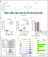
Genome-wide analysis and comprehensively characterization of somatic non-coding regulatory mutations in T-ALL. (A) Hotspot analysis identified loci with significant enrichment of recurrent noncoding mutations (single nucleotide variants and/or short insertion/deletions). Statistical significance (−log10 [P value], y-axis) is plotted against the respective chromosomal position of each mutation hotspot (x-axis). Dashed horizontal line, genome-wide significance threshold (P = 5 × 10⁻⁸); open circles, mutations significantly associated with expression of adjacent genes (TAL1, LMO1); dots below y = 1, sites with only 1 mutation. (B) Association of LMO1 enhancer mutation (11:8289481G>A) with LMO1 overexpression in the discovery cohort. FPKM, fragments per kilobase of transcript per million mapped reads. (C) Patients with TAL1 enhancer mutations (Mut) had the highest TAL1 expression compared with those with an intrachromosomal rearrangement (STIL-TAL1) or those with a wild-type (WT) genotype. (D) Among a panel of T-ALL cell lines, Jurkat cells were identified as having the LMO1 enhancer mutation and LMO1 overexpression. Gray and blue rectangles, T-ALL cases or cell lines with or without the LMO1 enhancer mutation, respectively. (E) All LMO1 enhancer mutation events were completely conserved across patients and created an MYB binding site. (F) In Jurkat cells, binding of MYB, CREBBP, and RUNX1 was detected by ChIP-seq, with the peak precisely superimposing the LMO1 enhancer mutation (red arrow). RNA polymerase II (RNAPII) binding, DNase I hypersensitivity, and H3K27 acetylation confirmed active transcription at this locus. ChIP-seq peak calling was performed using the MACS2 algorithm (enrichment P < 10−7). (G) Further analysis of the ChIP-seq reads of each allele indicates that transcription factor binding and transcription activation were specific to the mutant allele. ChIP-seq and DNase I hypersensitivity signals (y-axis) were normalized to reads per million reads sequenced in each sample. Statistical significance of the differences was estimated by using 2-sided Mann-Whitney-Wilcoxon test (B-C).
Figure 2.
Figure 2.
LMO1 enhancer mutation analyses for transcription activity in vitro and leukemia progression in vivo. (A) Jurkat cells were transfected with a reporter gene construct with WT sequence or LMO1 enhancer mutation, and transcription activation was quantified by luciferase activity relative to Renilla. LMO1 mutant enhancer activity was significantly diminished upon downregulation of MYB by short interfering RNA (siRNA). (B) In HEK293T cells, LMO1 enhancer mutation drove luciferase transcription only when MYB was ectopically expressed. (C) LMO1 overexpression was abrogated upon conversion of the LMO1 mutant allele to the WT allele in Jurkat cells, using CRISPR/Cas9 genomic engineering. (D) Engineered Jurkat cells with WT genotype at the LMO1 enhancer locus also showed a significantly lower rate of proliferation compared with parental Jurkat cells. (E) Jurkat cells were transduced with lentiviral vectors encoding constitutive Cas9 and isgLMO1. Parental Jurkat or CRISPR/Cas9-engineered cells were injected into sublethally irradiated NOD.Cg-PrkdcscidII2rgtm1Wjl/SzJ recipient mice. Mice bearing genome-edited Jurkat cells were split into 2 groups: with vs without doxycycline (Dox) treatment (ie, with vs without LMO1 enhancer deletion). (F) Leukemia progress was monitored weekly by complete blood count and flow cytometry quantification of human CD45 cells in peripheral blood. Median survival for mice bearing parental cells or isgLMO1 cells without doxycycline treatment was 29.5 and 32.5 days, respectively, whereas doxycycline-treated mice bearing isgLMO1 cells remained alive at the end of this experiment. Statistical significance of the differences was estimated by using 2-sided Mann-Whitney-Wilcoxon test (A-C, E), 2-way analysis of variance (D), or log-rank test (F); *P < .05, **P < .01, and ***P < .001. NS, not significant.

Comment in

References

    1. Roberts KG, Mullighan CG. Genomics in acute lymphoblastic leukaemia: insights and treatment implications. Nat Rev Clin Oncol. 2015;12(6):344-357. - PubMed
    1. Pui CH, Yang JJ, Hunger SP, et al. . Childhood acute lymphoblastic leukemia: progress through collaboration. J Clin Oncol. 2015;33(27):2938-2948. - PMC - PubMed
    1. Inaba H, Greaves M, Mullighan CG. Acute lymphoblastic leukaemia. Lancet. 2013;381(9881):1943-1955. - PMC - PubMed
    1. Belver L, Ferrando A. The genetics and mechanisms of T cell acute lymphoblastic leukaemia. Nat Rev Cancer. 2016;16(8):494-507. - PubMed
    1. Aifantis I, Raetz E, Buonamici S. Molecular pathogenesis of T-cell leukaemia and lymphoma. Nat Rev Immunol. 2008;8(5):380-390. - PubMed

Publication types