Review
. 2017 Apr 15;45(2):313-322.
doi: 10.1042/BST20160145.

The sweet side of the cell cycle

Affiliations
Review

The sweet side of the cell cycle

Ee Phie Tan et al. Biochem Soc Trans. .

Abstract

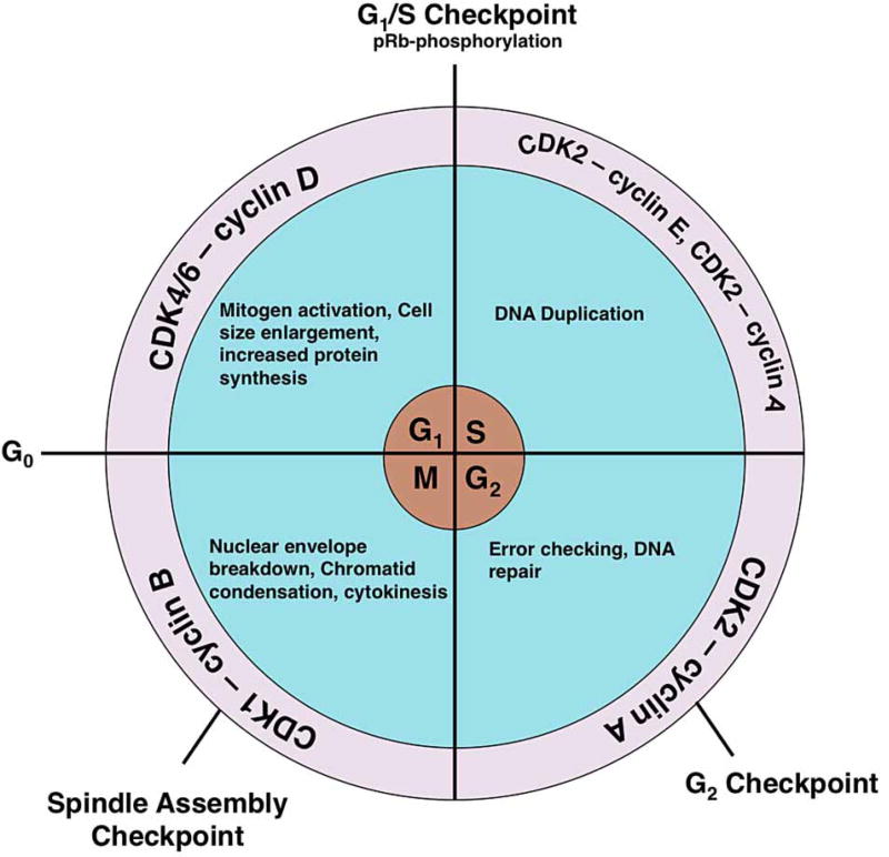
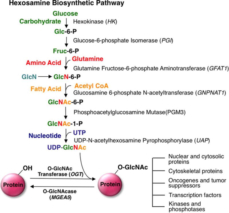
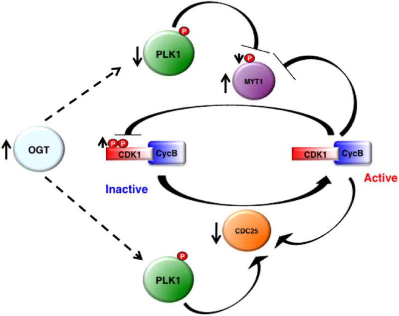
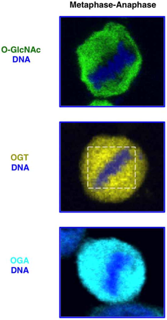
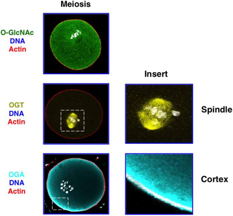
Cell division (mitosis) and gamete production (meiosis) are fundamental requirements for normal organismal development. The mammalian cell cycle is tightly regulated by different checkpoints ensuring complete and precise chromosomal segregation and duplication. In recent years, researchers have become increasingly interested in understanding how O-GlcNAc regulates the cell cycle. The O-GlcNAc post-translation modification is an O-glycosidic bond of a single β-N-acetylglucosamine sugar to serine/threonine residues of intracellular proteins. This modification is sensitive toward changes in nutrient levels in the cellular environment making O-GlcNAc a nutrient sensor capable of influencing cell growth and proliferation. Numerous studies have established that O-GlcNAcylation is essential in regulating mitosis and meiosis, while loss of O-GlcNAcylation is lethal in growing cells. Moreover, aberrant O-GlcNAcylation is linked with cancer and chromosomal segregation errors. In this review, we will discuss how O-GlcNAc controls different aspects of the cell cycle with a particular emphasis on mitosis and meiosis.

Keywords: O-GlcNAc; OGA; OGT; cell cycle; meiosis; mitosis.


Conflict of interest statement

Competing Interests

The Authors declare that there are no competing interests associated with the manuscript.

Figures

Figure 1
Figure 1. After mitogen activation, quiescent cells enter the cell cycle from the G0 phase
During the G1 phase, activation of CDK4/6 by cyclin D causes the cell size to increase, induces transcription of cell cycle genes, and leads to organelle duplication. The phosphorylation of the pRb protein promotes escape from the G1/S checkpoint and into the S-phase. Activation of CDK2 by cyclin E induces DNA synthesis, whereas CDK2 activation by cyclin A leads to the completion of DNA synthesis and promotes entry into the G2 phase. After the cell checks for error-free DNA replication, activation of CDK1 by cyclin B promotes M-phase entry. At the M-phase, the nuclear envelope breaks down, chromatids condense, the spindle forms, and the cell separates at cytokinesis into two daughter cells.
Figure 2
Figure 2. Schematic representation of the HBP and O-GlcNAc modification
Glutamine:fructose-6-phosphate (GFAT) is the rate-limiting step of the HBP pathway. GlcN treatment can bypass GFAT, causing an elevation in O-GlcNAcylation. The HBP is composed of various metabolic inputs including glucose, amino acid, fatty acid, and nucleotide metabolisms that ultimately serve to synthesize the donor substrate for OGT, UDP-GlcNAc. These metabolic inputs make O-GlcNAc a nutrient sensor capable of influencing many cellular processes including transcription, cell growth, and proliferation. Glc-6-P, glucose-6-phosphate; Fruc-6-P, fructose-6-phosphate; GlcN-6-P, glucosamine-6-phosphate; GlcNAc-6-P, N-acetylglucosamine-6-P; GlcNAc-1-P, N-acetylglucosamine-1-P. Arrow indicates that multiple steps are involved in the conversion.
Figure 3
Figure 3. CDK1 signaling is affected by elevations of O-GlcNAcylation
Overexpression of OGT causes PLK1 expression to decrease. Reduced PLK1 leads to decreased MYT1 phosphorylation and increased protein expression. Subsequently, elevation of MYT1 expression increases CDK1 inhibitory phosphorylation. Furthermore, CDC25 (CDK1 phosphatase) that is activated by PLK1 has a lower mRNA level after OGT overexpression.
Figure 4
Figure 4. In HeLA cells during metaphase–anaphase, a subset of OGT localizes to the spindle (white box) as determined by confocal microscopy
OGA and total cellular O-GlcNAc show no specific metaphase–anaphase localization. DNA (blue), O-GlcNAc (green), OGT (yellow), and OGA (cyan).
Figure 5
Figure 5. O-GlcNAc machinery localizes to discrete locations in mouse oocytes at the metaphase of meiosis I
OGT localizes to the meiotic spindle (see enlarged insert), whereas a subset of OGA localizes to the cell cortex (see enlarged insert). Images were taken using confocal microscopy. O-GlcNAc (green), OGT (yellow), OGA (cyan), and actin (red).

References

    1. Nurse P, Masui Y, Hartwell L. Understanding the cell cycle. Nat. Med. 1998;4:1103–1106. - PubMed
    1. Satyanarayana A, Kaldis P. Mammalian cell-cycle regulation: several Cdks, numerous cyclins and diverse compensatory mechanisms. Oncogene. 2009;28:2925–2939. - PubMed
    1. Murray AW. Recycling the cell cycle: cyclins revisited. Cell. 2004;116:221–234. - PubMed
    1. Slawson C, Copeland RJ, Hart GW. O-GlcNAc signaling: a metabolic link between diabetes and cancer? Trends Biochem. Sci. 2010;35:547–555. - PMC - PubMed
    1. Hart GW, Slawson C, Ramirez-Correa G, Lagerlof O. Cross talk between O-GlcNAcylation and phosphorylation: roles in signaling, transcription, and chronic disease. Annu. Rev. Biochem. 2011;80:825–858. - PMC - PubMed

Publication types