Review
. 2017 Apr 15;45(2):571-582.
doi: 10.1042/BST20160182.

Iminosugar antivirals: the therapeutic sweet spot

Affiliations
Review

Iminosugar antivirals: the therapeutic sweet spot

Dominic S Alonzi et al. Biochem Soc Trans. .

Abstract

Many viruses require the host endoplasmic reticulum protein-folding machinery in order to correctly fold one or more of their glycoproteins. Iminosugars with glucose stereochemistry target the glucosidases which are key for entry into the glycoprotein folding cycle. Viral glycoproteins are thus prevented from interacting with the protein-folding machinery leading to misfolding and an antiviral effect against a wide range of different viral families. As iminosugars target host enzymes, they should be refractory to mutations in the virus. Iminosugars therefore have great potential for development as broad-spectrum antiviral therapeutics. We outline the mechanism giving rise to the antiviral activity of iminosugars, the current progress in the development of iminosugar antivirals and future prospects for this field.

Keywords: calnexin; drug discovery and design; glucosidase; glycobiology; iminosugar.


Conflict of interest statement

The Authors declare that there are no competing interests associated with the manuscript.

Figures
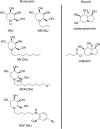
Figure 1.
Figure 1.. Structures of iminosugars discussed in this mini-review.
The left panel shows the structure of the naturally occurring iminosugar DNJ, along with derivatives modified to increase antiviral efficacy. MON-DNJ, currently in clinical trial against dengue, is also referred to as UV4B. The right panel shows the structure of the naturally occurring iminosugar CAST along with the prodrug celgosivir which is in a phase II clinical trial against dengue.
Figure 2.
Figure 2.. The calnexin cycle and ERAD.
The precursor glycan Glc3Man9GlcNAc2 (represented here for simplicity with the glucose residues as red triangles and the remaining portion of the glycan shown as black lines) is added to a peptide co-translationally. Cleavage of the terminal glucose residue by α-glu I leads to a form that can either bind to malectin or be further trimmed by α-glu II to become a substrate for calnexin/calreticulin. On release from calnexin/calreticulin, α-glu II can remove the remaining glucose residue. At this point properly folded proteins are exported to the Golgi for further processing, whilst misfolded proteins are either reglucosylated by UGGT for a ‘second chance’ at folding or directed to the ERAD pathway by ER mannosidase I (ER Man I), which removes a mannose residue from the B-arm of the glycan [42,79]. ER degradation-enhancing α-mannosidase-like proteins 1–3 (EDEM1–3) then act on the C-arm of the glycan followed by OS-9/XTP3-B-mediated delivery of the substrate to the Hrd1 ubiquitination complex through the interaction with a membrane-spanning adaptor protein, SEL1L [–87]. PNGase separates the glycan from the protein and both segments are degraded [44,88].
Figure 3.
Figure 3.. FOS analysis of cells grown in the presence of iminosugars.
(A) FOS are produced by the activity of two PNGase enzymes: one located in the ER, and the other in the cytosol. In the absence of iminosugar inhibitors FOS produced in the ER will be exported via a FOS transporter to the cytosol for degradation. The presence of terminal glucose residues on the A-arm of the glycan prevents export from the ER, leading to an increase in glucosylated FOS in the ER in the presence of iminosugars. Misfolded glycoproteins targeted for degradation through ERAD are trimmed by the enzymes ENGase and a cytosolic mannosidase. In the absence of iminosugars the resulting glycans enter the lysosome for further degradation; however, the presence of glucose residues prevents this leading to a build-up of glucosylated FOS in the cytosol. (B) Following isolation, purification and fluorescent labelling, total cellular FOS containing both the ER pool (1 in A) and cytosolic FOS pool (2 in A) can be analysed by NP-HPLC. Shown here are representative chromatograms of total cellular FOS from control HL60s cells (upper panel) and HSL60s cells treated with 1 mM NB-DNJ for 24 h (lower panel). The glucose capped glycans visible in the lower panel are indicative of α-glucosidase inhibition.
Figure 4.
Figure 4.. The structure of α-glu II; active site and in complex with inhibitors.
(A) Ribbon diagram illustrating the structure of α-glu II with the key catalytic residues shown as sticks. The catalytic subunit is shown in gold, whilst the fragment of the accessory subunit remaining after protease digestion is shown in silver. (B) The catalytic binding site of α-glu II. Key residues for catalytic activity and interaction with the substrate glycan are shown as sticks. In the catalytic cycle D564 acts as a nucleophile and D640 as the catalytic acid/base. As is typical for sugar-binding sites, there are many aromatic residues and polar groups offering a wide range of potential interactions for inhibitors. (C) The catalytic binding site of α-glu II with NB-DNJ shown in magenta as a ball-and-stick representation. In this structure, the glucose-like portion occupies a similar position to the natural substrate and the alkyl chain occupies the +1 site, which would be occupied by the sugar moiety adjacent to that cleaved in the reaction in the natural substrate. The side chain of W525 becomes disordered and is not shown in the structure. (D) The catalytic binding site of α-glu II with the inhibitor MON-DNJ shown in purple as a ball-and-stick representation. In this structure, the alkyl chain of the iminosugar fully occupies the +1 site and extends beyond; the alkyl chain adopts two main conformations, one of which is located towards the +2 sugar-binding site. Addition of the inhibitor leads to disorder in the catalytic site including displacement of the 523–528 loop (containing W525) and multiple conformations for F571. In all figures, H atoms are removed for clarity and all structures are shown in the same orientation. Protein databank file 5foe.pdb was shown in A and B; 5ieg.pdb and 5ief.pdb were used for C and D, respectively.

References

    1. Block T.M., Lu X., Platt F.M., Foster G.R., Gerlich W.H., Blumberg B.S. et al. (1994) Secretion of human hepatitis B virus is inhibited by the imino sugar N-butyldeoxynojirimycin. Proc. Natl Acad. Sci. U.S.A. 91, 2235–2239 doi:10.1073/pnas.91.6.2235 - DOI - PMC - PubMed
    1. Hammond C., Braakman I. and Helenius A. (1994) Role of N-linked oligosaccharide recognition, glucose trimming, and calnexin in glycoprotein folding and quality control. Proc. Natl Acad. Sci. U.S.A. 91, 913–917 doi:10.1073/pnas.91.3.913 - DOI - PMC - PubMed
    1. Mehta A., Lu X., Block T.M., Blumberg B.S. and Dwek R.A. (1997) Hepatitis B virus (HBV) envelope glycoproteins vary drastically in their sensitivity to glycan processing: evidence that alteration of a single N-linked glycosylation site can regulate HBV secretion. Proc. Natl Acad. Sci. U.S.A. 94, 1822–1827 doi:10.1073/pnas.94.5.1822 - DOI - PMC - PubMed
    1. Dwek R.A., Butters T.D., Platt F.M. and Zitzmann N. (2002) Targeting glycosylation as a therapeutic approach. Nat. Rev. Drug Discovery 1, 65–75 doi:10.1038/nrd708 - DOI - PubMed
    1. Chang J., Guo J.-T., Du Y. and Block T. (2013) Imino sugar glucosidase inhibitors as broadly active anti-filovirus agents. Emerg. Microbes Infect. 2, e77 doi:10.1038/emi.2013.77 - DOI - PMC - PubMed

MeSH terms