Review
. 2017 Aug;38(8):1467-1474.
doi: 10.3174/ajnr.A5138. Epub 2017 Apr 13.

Dentate Update: Imaging Features of Entities That Affect the Dentate Nucleus

Affiliations
Review

Dentate Update: Imaging Features of Entities That Affect the Dentate Nucleus

K M Bond et al. AJNR Am J Neuroradiol. 2017 Aug.

Abstract

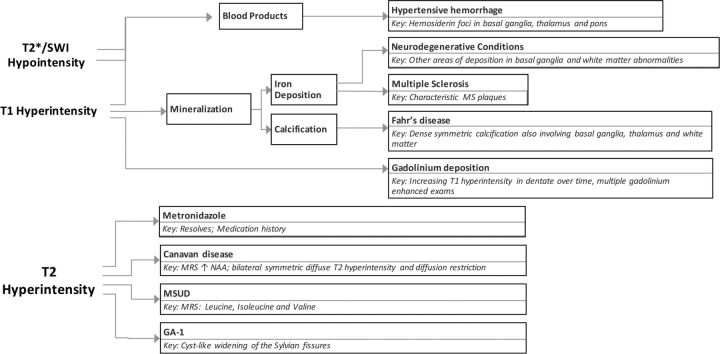
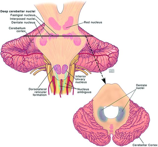
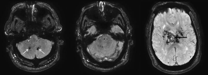
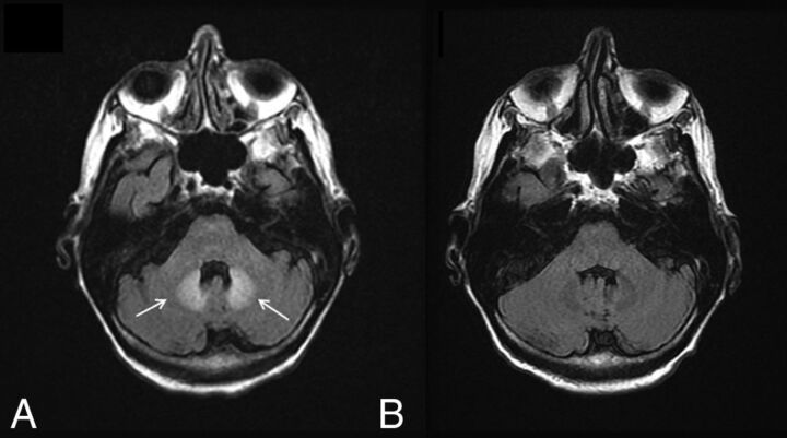
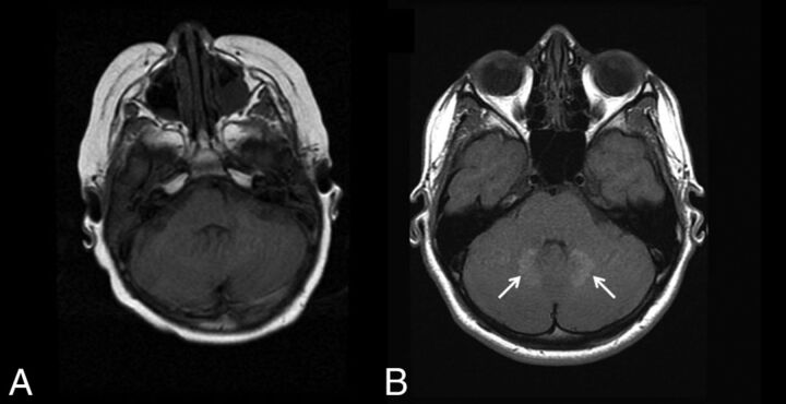
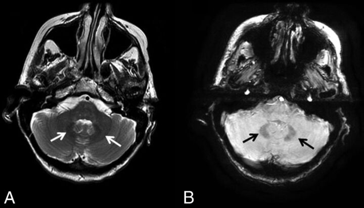
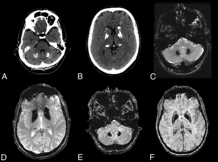
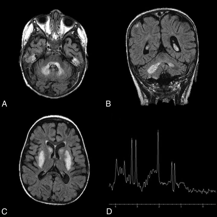
The dentate nucleus is a cerebellar structure involved in voluntary motor function and cognition. There are relatively few entities that affect the dentate, and the clinical features of these conditions are often complex and nonspecific. Because these entities are rarely encountered, the formulation of a differential diagnosis can be difficult. Many of the conditions are reversible or treatable with early intervention. Therefore, it is important to recognize classic clinical presentations and their associated characteristic imaging findings. We provide a summary of entities that affect the dentate nucleus and a diagnostic workflow for approaching dentate nucleus imaging abnormalities.


Figures

Fig 1.
Fig 1.
Diagnostic approach for dentate nucleus imaging abnormalities.
Fig 2.
Fig 2.
Illustration of the location of the dentate nucleus within the cerebellum. Used with permission of Mayo Foundation for Medical Education and Research, all rights reserved.
Fig 3.
Fig 3.
Hypertensive hemorrhage. A 55-year-old man who presented with a thalamic hemorrhage. Three selected images from 3D susceptibility-weighted imaging demonstrate microhemorrhages and hemosiderin deposition within the dentate nuclei (white arrows), brain stem, thalamus, and basal ganglia. The sentinel bleed is shown with black arrows. This distribution is characteristic of hypertensive microhemorrhages. The patient was being treated with multiple medications to control his hypertension, which remained elevated even during his hospitalization.
Fig 4.
Fig 4.
Metronidazole toxicity. A 74-year-old woman was treated for 6 weeks with metronidazole and developed dysarthria. T2 FLAIR images demonstrate T2 hyperintensity within the dentate nuclei (A, arrows), which resolved on MR imaging 1 month later (B).
Fig 5.
Fig 5.
Gadolinium deposition. A 13-year-old boy was diagnosed with a pilocytic astrocytoma and treated with chemotherapy, but no radiation. A, the first available pregadolinium T1WI without any abnormality in the dentate. B, A follow-up examination performed 10 years later now demonstrates T1 hyperintensity on TIWI within the dentate nuclei (arrows). The patient had undergone 37 gadolinium-enhanced examinations at this time point.
Fig 6.
Fig 6.
Neurodegeneration with brain iron accumulation. A 40-year-old woman with Woodhouse-Sakati syndrome found to have neurodegenerative iron accumulation. Marked hypointensity within the dentate on T2WI (A, white arrows) and 3D SWI (B, black arrows) is consistent with iron deposition.
Fig 7.
Fig 7.
Fahr disease. A–D, CT and T2* gradient recalled-echo images from a 49-year-old patient with Fahr disease demonstrate dense calcification of the dentate nuclei and basal ganglia. E and F, Dense calcification within the dentate nuclei and basal ganglia is also well-demonstrated on susceptibility-weighted imaging, in this case from a 68-year-old affected woman.
Fig 8.
Fig 8.
Leigh disease. An 11-year-old girl diagnosed at 2 years of age with Leigh disease. Marked T2 hyperintensity on FLAIR within the dentate nuclei (A and B) and basal ganglia (C). MR spectroscopy with voxel sampling of the basal ganglia (D) demonstrates a prominent lactate doublet (at 1.3 ppm) and elevated choline (at 3.2 ppm).

References

    1. Rhoton AL Jr. Cerebellum and fourth ventricle. Neurosurgery 2000;47:S7–27 10.1097/00006123-200009001-00007 - DOI - PubMed
    1. Akakin A, Peris-Celda M, Kilic T, et al. . The dentate nucleus and its projection system in the human cerebellum: the dentate nucleus microsurgical anatomical study. Neurosurgery 2014;74:401–24; discussion 424–25 10.1227/NEU.0000000000000293 - DOI - PubMed
    1. O'Halloran CJ, Kinsella GJ, Storey E. The cerebellum and neuropsychological functioning: a critical review. J Clin Exp Neuropsychol 2012;34:35–56 10.1080/13803395.2011.614599 - DOI - PubMed
    1. Schmahmann JD, Sherman JC. The cerebellar cognitive affective syndrome. Brain 1998;121(pt 4):561–79 10.1093/brain/121.4.561 - DOI - PubMed
    1. Timmann D, Drepper J, Frings M, et al. . The human cerebellum contributes to motor, emotional and cognitive associative learning: a review. Cortex 2010;46:845–57 10.1016/j.cortex.2009.06.009 - DOI - PubMed

LinkOut - more resources