. 2017 Jun 1;312(6):G649-G657.
doi: 10.1152/ajpgi.00366.2016. Epub 2017 Apr 13.

Gastrin induces parathyroid hormone-like hormone expression in gastric parietal cells

Affiliations

Gastrin induces parathyroid hormone-like hormone expression in gastric parietal cells

Asma Al Menhali et al. Am J Physiol Gastrointest Liver Physiol. .

Abstract

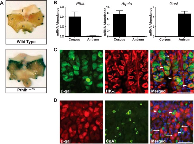
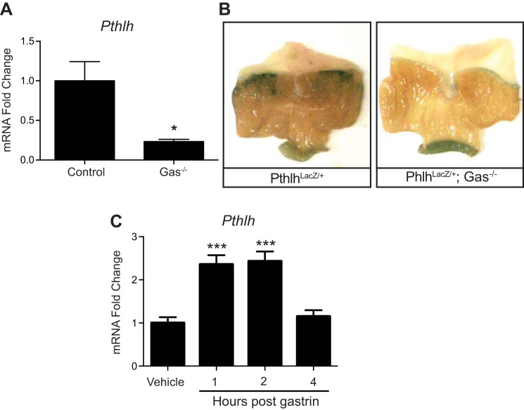
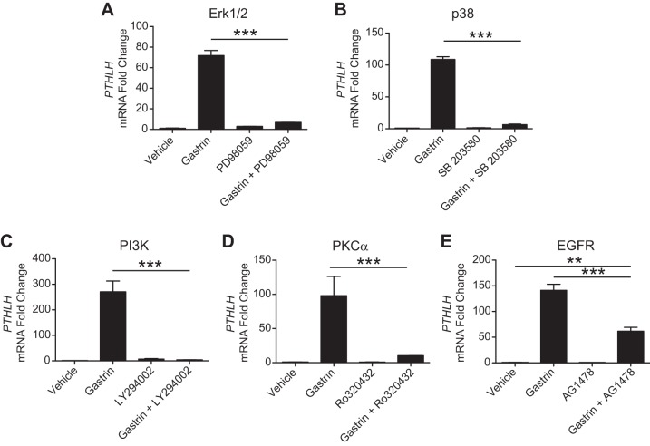
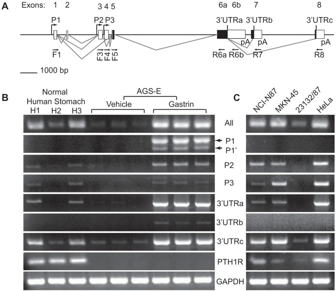
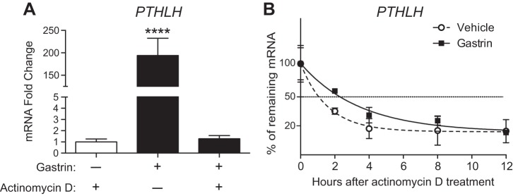
Parietal cells play a fundamental role in stomach maintenance, not only by creating a pathogen-free environment through the production of gastric acid, but also by secreting growth factors important for homeostasis of the gastric epithelium. The gastrointestinal hormone gastrin is known to be a central regulator of both parietal cell function and gastric epithelial cell proliferation and differentiation. Our previous gene expression profiling studies of mouse stomach identified parathyroid hormone-like hormone (PTHLH) as a potential gastrin-regulated gastric growth factor. Although PTHLH is commonly overexpressed in gastric tumors, its normal expression, function, and regulation in the stomach are poorly understood. In this study we used pharmacologic and genetic mouse models as well as human gastric cancer cell lines to determine the cellular localization and regulation of this growth factor by the hormone gastrin. Analysis of PthlhLacZ/+ knock-in reporter mice localized Pthlh expression to parietal cells in the gastric corpus. Regulation by gastrin was demonstrated by increased Pthlh mRNA abundance after acute gastrin treatment in wild-type mice and reduced expression in gastrin-deficient mice. PTHLH transcripts were also observed in normal human stomach as well as in human gastric cancer cell lines. Gastrin treatment of AGS-E gastric cancer cells induced a rapid and robust increase in numerous PTHLH mRNA isoforms. This induction was largely due to increased transcriptional initiation, although analysis of mRNA half-life showed that gastrin treatment also extended the half-life of PTHLH mRNA, suggesting that gastrin regulates expression by both transcriptional and posttranscriptional mechanisms.NEW & NOTEWORTHY We show that the growth factor parathyroid hormone-like hormone (PTHLH) is expressed in acid-secreting parietal cells of the mouse stomach. We define the specific PTHLH mRNA isoforms expressed in human stomach and in human gastric cancer cell lines and show that gastrin induces PTHLH expression via transcription activation and mRNA stabilization. Our findings suggest that PTHLH is a gastrin-regulated growth factor that might contribute to gastric epithelial cell homeostasis.

Keywords: AU-rich element; PTHrP; growth factor; mRNA stability; parathyroid hormone-related protein; stomach.


Figures

Fig. 1.
Fig. 1.
Gastric parietal cells express parathyroid hormone-like hormone (Pthlh). A: whole mount 5-bromo-4-chloro-3-indyl-β-d-galactosidase (X-gal) staining in PthlhLacZ/+ (bottom) and wild-type (top) mouse stomach localized LacZ reporter expression to the corpus region (f, forestomach; c, corpus; a, antrum). The dark edge staining is an artifact due to pinning the tissue. B: quantitative (q)RT-PCR measurement of mRNA abundance for parathyroid hormone-like hormone (Pthlh), the corpus marker H+-K+-ATPase α-subunit (Atp4a), and the antral marker gastrin (Gast) in total RNA isolated from mouse gastric corpus and antrum. Values were normalized to Gapdh and are presented as means ± SE (n = 3–5). C: coimmunostaining PthlhLacZ/+ stomach cryosections with antibodies against the reporter β-galactosidase (β-gal; green) and the parietal cell marker H+-K+-ATPase α-subunit (HK-α; red) showed colocalization in a subset of parietal cells. Arrowheads, β-gal-expressing parietal cells; arrow, non-β-gal-expressing parietal cell. D: coimmunostaining of β-gal (red) and the enterochromaffin-like marker chromogranin A (CgA; green) showed no colocalization. Arrowheads, CgA-expressing cells; arrows, β-gal-expressing cells. Scale bars = 50 µm.
Fig. 2.
Fig. 2.
Gastrin stimulates Pthlh expression in vivo. A: Pthlh mRNA abundance was measured by qRT-PCR analysis of corpus RNA isolated from gastrin-deficient mice (Gas−/−) and Gas+/+ control littermates. *P < 0.05 vs. control by Student’s t-test. B: whole mount X-gal staining of PthlhLacZ/+ reporter mouse stomach on control or Gas−/− strain background. Staining at the pylorus is nonspecific. C: qRT-PCR measurement of Pthlh mRNA abundance in corpus isolated from wild-type mice at various times after gastrin injection (250 μg/kg). ***P < 0.001 vs. vehicle-injected group by a one-way ANOVA with Dunnett’s posttest. For A and C, data (means ± SE) are shown in reference to Gapdh expression measured in the same samples (n = 4 mice per group).
Fig. 3.
Fig. 3.
Gastrin stimulates PTHLH expression in vitro. A: qRT-PCR measurement of PTHLH (left) or TFF2 (right) mRNA abundance in AGS-E cells treated with vehicle or human gastrin (10−7 M). **P < 0.005, ****P ≤ 0.0001 vs. time 0 for each treatment by two-way ANOVA with Dunnett’s posttest. B: qRT-PCR measurement of PTHLH mRNA abundance in AGS and AGS-E cells 6 h after gastrin administration. ***P ≤ 0.0005 vs. all groups by two-way ANOVA with Tukey’s posttest. All data (means ± SE) are shown in reference to GAPDH expression measured in the same samples (n = 3 independent RNA samples per group).
Fig. 4.
Fig. 4.
Signaling pathways mediating gastrin induction of PTHLH. Pharmacological agents were used to inhibit various signaling pathways downstream of the gastrin receptor CCKBR. A: Erk1/2 inhibition with PD98059 (5 × 10⁻⁷ M). B: p38 inhibition with SB203580 (10−5 M). C: PI3K inhibition with LY294002 (1.5 × 10⁻⁵ M). D: PKCa inhibition with Ro320432 (10−6 M). E: EGFR inhibition with AG1478 (9 × 10⁻⁶ M). qRT-PCR measurement of PTHLH mRNA abundance in vehicle-treated, gastrin-treated, inhibitor-pretreated, or inhibitor-pretreated and gastrin-treated AGS-E cells 4 h after treatment. Values were normalized to GAPDH, and data are shown as means ± SE. **P < 0.01, ***P < 0.001, via one-way ANOVA with Tukey’s posttest (N = 3 independent RNA samples per group).
Fig. 5.
Fig. 5.
Gastrin induces multiple PTHLH mRNA isoforms. A: diagram of human PTHLH gene structure. The PTHLH gene consists of 8 exons (untranslated, white; translated, black) with 3 transcriptional starts (P1, P2, and P3). Alternative splicing generates isoforms with different 3′-untranslated regions (3′-UTRs: a, b, and c). Primer sets were designed to amplify all PTHLH mRNA species or specific isoforms from promoters P1, P2, and P3 and the 3′-UTRs (a, b, or c; see Tables 2 and 3 for primer sequences and amplicon information). The positions of the forward (F) and reverse (R) primers are indicated below the gene diagram. B: RT-PCR products generated from the RNA samples from normal human stomach (H1, H2, and H3) and RNAs from vehicle- or gastrin-treated AGS-E cells. RNAs were tested for the various indicated PTHLH isoforms and PTHLH receptor (PTH1R) expression; GAPDH was used as a reference for RNA quality and quantity. C: RT-PCR analysis of human gastric cancer cell lines as indicated. HeLa cells were used as a positive control.
Fig. 6.
Fig. 6.
Gastrin promotes PTHLH transcription and stabilizes PTHLH mRNA. A: analysis of PTHLH expression in AGS-E cells pretreated with actinomycin D 30 min before gastrin administration. mRNA abundance was measured 4 h postgastrin or vehicle administration. Values were normalized to GAPDH, and data are shown as means ± SE. ****P < 0.0001, via one-way ANOVA with Tukey’s posttest (n = 3 independent RNA samples per group). B: analysis of PTHLH mRNA half-life in AGS-E cells treated with vehicle or gastrin. Actinomycin D was added 4 h after gastrin induction and PTHLH mRNA abundance was measured at varying times by qRT-PCR analysis. Values were normalized to GAPDH and plotted to compare with values at time 0 (100%) when actinomycin D was applied.

References

    1. Alipov GK, Ito M, Nakashima M, Ikeda Y, Nakayama T, Ohtsuru A, Yamashita S, Sekine I. Expression of parathyroid hormone-related peptide (PTHrP) in gastric tumours. J Pathol 182: 174–179, 1997. doi: 10.1002/(SICI)1096-9896(199706)182:2<174::AID-PATH840>3.0.CO;2-4. - DOI - PubMed
    1. Andersson N, Skrtic SM, Håkanson R, Ohlsson C. A gene expression fingerprint of mouse stomach ECL cells. Biochem Biophys Res Commun 332: 404–410, 2005. doi: 10.1016/j.bbrc.2005.05.004. - DOI - PubMed
    1. Beales IL. Gastrin and interleukin-1beta stimulate growth factor secretion from cultured rabbit gastric parietal cells. Life Sci 75: 2983–2995, 2004. doi: 10.1016/j.lfs.2004.03.041. - DOI - PubMed
    1. Chen X, Macica CM, Dreyer BE, Hammond VE, Hens JR, Philbrick WM, Broadus AE. Initial characterization of PTH-related protein gene-driven lacZ expression in the mouse. J Bone Miner Res 21: 113–123, 2006. doi: 10.1359/JBMR.051005. - DOI - PubMed
    1. Cho YM, Lewis DA, Koltz PF, Richard V, Gocken TA, Rosol TJ, Konger RL, Spandau DF, Foley J. Regulation of parathyroid hormone-related protein gene expression by epidermal growth factor-family ligands in primary human keratinocytes. J Endocrinol 181: 179–190, 2004. doi: 10.1677/joe.0.1810179. - DOI - PubMed

MeSH terms