. 2017 Apr 14:23:1800-1811.
doi: 10.12659/msm.902754.

Glial Cell Line-Derived Neurotrophic Factor-Transfected Placenta-Derived Versus Bone Marrow-Derived Mesenchymal Cells for Treating Spinal Cord Injury

Affiliations

Glial Cell Line-Derived Neurotrophic Factor-Transfected Placenta-Derived Versus Bone Marrow-Derived Mesenchymal Cells for Treating Spinal Cord Injury

Yao Lu et al. Med Sci Monit. .

Abstract

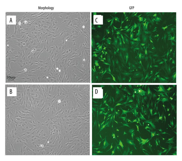
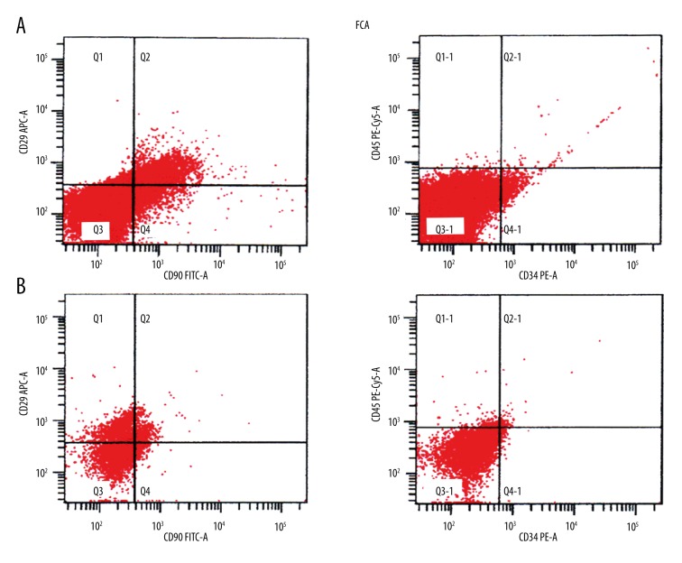
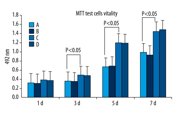
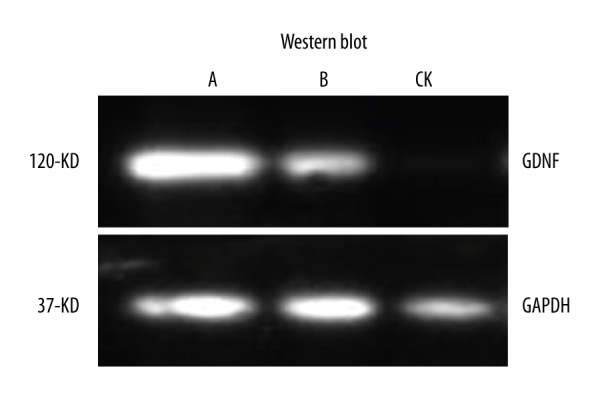
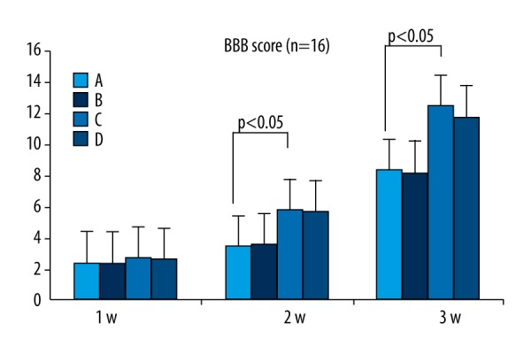
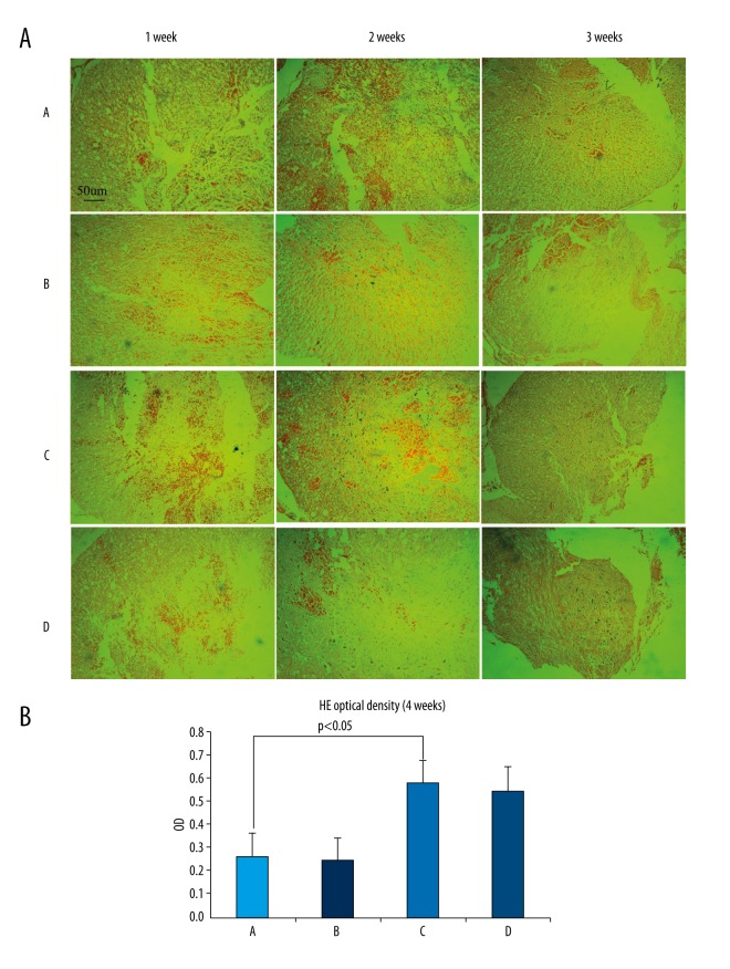
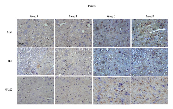
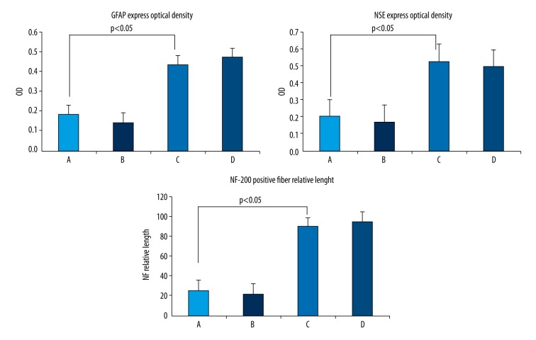
BACKGROUND Placenta-derived mesenchymal stem cells (PMSCs) were isolated from placenta and had differentiation and self-renewal potential. We transfected PMSCs with glial cell line-derived neurotrophic factor (GDNF) and compared their effect for repairing spinal cord injury (SCI) with that of GDNF-transfected bone marrow-derived mesenchymal stem cell (BMSC). MATERIAL AND METHODS The PMSCs were isolated from Sprague-Dawley rat placenta; BMSCs were isolated from Sprague-Dawley rat thigh bone marrow. Primary cultured BMSCs and PMSCs were uniformly spindle-shaped. Flow cytometry indicated that both cell types were CD29- and CD90-positive and CD34- and CD45-negative, confirming that they were MSCs. The PMSCs and BMSCs were transfected with recombinant lentivirus containing the GDNF gene in vitro. PMSC and BMSC viability was increased after transfection, and GDNF expression was increased until 10 d after transfection. SCI was created in the rats (n=64) and was repaired using transfected PMSCs and BMSCs or untransfected PMSCs and BMSCs. RESULTS The transfected PMSCs and BMSCs repaired the SCI. Flow cytometry, histology, immunohistochemical, kinesiology properties, and Basso-Beattie-Bresnahan locomotion score measurements determined no significant difference between transfected PMSCs and BMSCs at 7, 14, and 21 d post-transplantation (P>0.05); the injury healed better in transfected PMSCs and BMSCs than in untransfected PMSCs and BMSCs (P<0.05). CONCLUSIONS MSCs have similar biology characteristics and capacity for SCI repair to BMSCs and can be used as a new resource for treating SCI.


Figures

Figure 1
Figure 1
(A, B) PMSC and BMSC morphology under inverted phase-contrast microscopy (×100). Day 5 first-generation PMSCs (A) and BMSCs (B) are elongated spindle-shaped and followed similar patterns such as uniform fibroblast-like colony growth. (C, D) Morphology of GDNF-transfected PMSCs and BMSCs. Cells were observed under inverted phase-contrast microscopy (×100), MOI=100 12 h after transfection. GFP expression of PMSCs (C) and BMSCs (D) was significant and the cell morphology was intact without obvious cytopathic effects.
Figure 2
Figure 2
Flow cytometry of PMSCs and BMSCs. CD29 and CD90 were highly expressed in PMSCs (A) and BMSCs (B), while CD34 and CD45 were not.
Figure 3
Figure 3
MTT assay of untransfected PMSCs (A) and untransfected BMSCs (B) and GDNF-transfected PMSCs (C) and GDNF-transfected BMSCs (D). After 3–5 d of transfection, PMSCs and BMSCs exhibited logarithmic growth and the growth rate slowed at 6–7 d. The cell viability of all 4 groups was not significantly different (P>0.05) at 24 h after transfection. At 3 d after transfection, the cell viability of GDNF-transfected-PMSCs and GDNF-transfected-BMSCs gradually became significantly higher than that of PMSCs and BMSCs (P<0.05). The cell viability of GDNF-transfected-PMSCs and GDNF-transfected-BMSCs was not significantly different (P > 0.05).
Figure 4
Figure 4
Western blotting showing that PMSCs (A) and BMSCs (B) express GDNF after 7-d GDNF transfection.
Figure 5
Figure 5
BBB score. At 1, 2, and 4 weeks after surgery, the BBB score between PMSCs (A) and BMSCs (B) and GDNF-transfected-PMSCs (C) and GDNF-transfected-BMSCs (D) were significantly different (P<0.05); the difference between GDNF-transfected-PMSCs (C) and GDNF-transfected-BMSCs (D) was not significant (P>0.05).
Figure 6
Figure 6
(A, B) HE staining observed under inverted phase-contrast microscopy (×100). One week after surgery, inflammatory cells, cavities, and nerve fiber necrosis can be observed in the SCI. Nerve cell proliferation can be also observed in GDNF-transfected-PMSCs (C) and GDNF-transfected-BMSCs (D). After 2 weeks and in all 4 groups, the SCI was filled with dense fibrous connective tissue. Astrocyte proliferation and accumulation in the glial scar is visible. There were obviously more proliferating cells in GDNF-transfected-PMSCs (C) and GDNF-transfected-BMSCs (D) than in PMSCs (A) and BMSCs (B). After 4 weeks, the scar tissue can still be observed in each group but with reduced cysts and necrosis. The tissue is denser, while there are significantly increased nerve cells as compared to that at 2 weeks. There was significantly increased nerve cell proliferation in GDNF-transfected-PMSCs (C) and GDNF-transfected-BMSCs (D) but it was much lower in PMSCs (A) and BMSCs (B). Overall, GDNF-transfected-PMSCs (C) and GDNF-transfected-BMSCs (D) did not differ greatly, but were significantly improved as compared to PMSCs (A) and BMSCs (B).
Figure 7
Figure 7
GFAP expression in the SCI area was significantly higher in GDNF-transfected-PMSCs (C) and GDNF-transfected-BMSCs (D) than in PMSCs (A) and BMSCs (B). The blue nuclei are surrounded by the brown-stained cytoplasm. NSE expression in the SCI area was significantly higher in GDNF-transfected-PMSCs (C) and GDNF-transfected-BMSCs(D) than in PMSCs (A) and BMSCs (B). The blue nuclei are surrounded by brown-stained cytoplasm. The NF-200 nerve fibers in the SCI area were significantly longer in GDNF-transfected-PMSCs (C) and GDNF-transfected-BMSCs (D) than in PMSCs (A) and BMSCs (B). GDNF-transfected-PMSCs (C) and GDNF-transfected-BMSCs (D) were not obviously different.
Figure 8
Figure 8
The optical density of GFAP expression in the SCI area was higher in GDNF-transfected-PMSCs (C) and GDNF-transfected-BMSCs (D) than in PMSCs (A) and BMSCs (B). The optical density of NSE expression in the SCI area was higher in GDNF-transfected-PMSCs (C) and GDNF-transfected-BMSCs (D) than in PMSCs (A) and BMSCs (B). The relative length of NF-200–positive nerve fibers in the SCI area was greater in GDNF-transfected-PMSCs (C) and GDNF-transfected-BMSCs (D) than in PMSCs (A) and BMSCs (B).

References

    1. Anderberg L, Aldskogius H, Holtz A. Spinal cord injury – scientific challenges for the unknown future. Ups J Med Sci. 2007;112:259–88. - PubMed
    1. Mitchell CS, Lee RH. Pathology dynamics predict spinal cord injury therapeutic success. J Neurotrauma. 2008;25:1483–97. - PMC - PubMed
    1. Bae JS, Han HS, Youn DH. Bone marrow-derived mesenchymal stem cells promote neuronal networks with functional synaptic transmission after transplantation into mice with neurodegeneration. Stem Cells. 2007;5:1307–16. - PubMed
    1. Dezawa M, Ishikawa H, Hoshino M. Potential of bone marrow stromal cells in applications for neuro-degenerative, neuro-traumatic and muscle degenerative disease. Curr Neuropharmacol. 2005;4:257–66. - PMC - PubMed
    1. Lu D, Li Y, Wang L. Intraarterial administration of marrow stromal cells in a rat model of traumatic brain injury. J Neurotrauma. 2001;8:813–19. - PubMed

MeSH terms

Substances