. 2017 Apr 13;7(1):880.
doi: 10.1038/s41598-017-00853-x.

MIEF1/2 function as adaptors to recruit Drp1 to mitochondria and regulate the association of Drp1 with Mff

Affiliations

MIEF1/2 function as adaptors to recruit Drp1 to mitochondria and regulate the association of Drp1 with Mff

Rong Yu et al. Sci Rep. .

Abstract

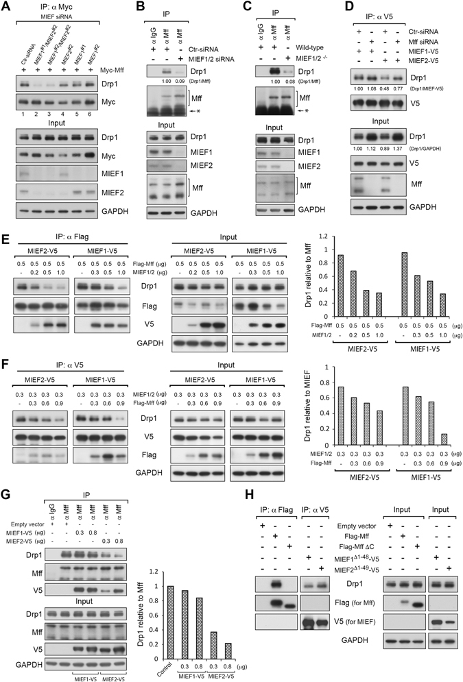
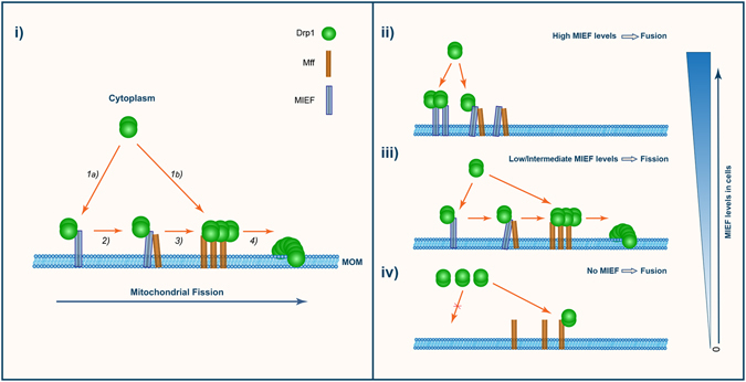
Mitochondrial dynamics is a fundamental cellular process and recruitment of Drp1 to mitochondria is an essential step in mitochondrial fission. Mff and MIEF1/2 (MiD51/49) serve as key receptors for recruitment of Drp1 to mitochondria in mammals. However, if and how these receptors work together in mitochondrial fission is poorly understood. Here we show that MIEFs interact with both Drp1 and Mff on the mitochondrial surface and serve as adaptors linking Drp1 and Mff together in a trimeric Drp1-MIEF-Mff complex. Thus, MIEFs can regulate the interaction between Drp1 and Mff, and also Mff-induced Drp1 accumulation on mitochondria. It is shown that loss of endogenous MIEFs severely impairs these processes. Additionally, in cells depleted of endogenous MIEF1/2, high levels of exogenous MIEFs sequester Drp1 on the mitochondrial surface, resulting in mitochondrial elongation, whereas low-to-moderate levels of MIEFs promote mitochondrial fission, leading to mitochondrial fragmentation. In sum, the data suggest that MIEFs and Mff work coordinately in Drp1-mediated mitochondrial fission and that the level of MIEF1/2 relative to Mff sets the balance between mitochondrial fission and fusion.


Conflict of interest statement

The authors declare that they have no competing interests.

Figures

Figure 1
Figure 1
MIEFs and Mff recruit Drp1 to mitochondria, but have opposing effects on mitochondrial morphology. (A) Overexpression of either MIEF1, MIEF2 or Mff recruits Drp1 from the cytoplasm to mitochondria, but MIEF overexpression leads to a mitochondrial fusion phenotype, while Mff induces mitochondrial fission. Confocal images of mitochondrial morphology and Drp1 distribution in 293T cells transfected with indicated plasmids, stained with MitoTracker (red), anti-Drp1 (green), and either anti-V5 (blue for MIEF) or anti-Myc (blue for Mff) antibodies. (B) Confocal images of 293T cells treated with control siRNA or a combination of MIEF1 and MIEF2 siRNAs, followed by transfection with empty vector or Myc-Mff plasmid, and stained with MitoTracker (red), anti-Drp1 (green) and anti-Myc (blue) antibodies. Insets represent high magnification views of the boxed areas. Results from quantitative co-localization of Drp1 with mitochondria are summarized in Fig. 3D.
Figure 2
Figure 2
Ablation of MIEF1/2 impairs Mff-induced accumulation of Drp1 on mitochondria, whereas knockdown of Mff does not impair MIEF-mediated recruitment of Drp1 to mitochondria. (A) Confocal images of mitochondrial morphology and Drp1 distribution in wild-type and MIEF1/2 DKO 293T cells transfected with empty vector or Myc-Mff plasmid. Cells were stained with MitoTracker (red) followed by immunostaining with anti-Drp1 (green) and anti-Myc (blue) antibodies. Insets represent high magnification views of the boxed areas. Quantitative co-localization of Drp1 with mitochondria is summarized in Fig. 3D. (B) Percentages (mean ± SEM) of cells with indicated mitochondrial morphologies in wild-type and MIEF1/2 DKO 293T cells transfected with empty vector or Myc-Mff plasmid in three independent experiments (≥150 cells were analyzed per experiment). (C) Knockdown of Mff does not affect MIEF-mediated recruitment of Drp1 to mitochondria. Confocal images show Drp1 distribution on mitochondria and in the cytoplasm. 293T cells were treated with control siRNA, or Mff siRNA, followed by transfection with either empty vector or with MIEF1-V5 or MIEF2-V5 as indicated, and stained with MitoTracker (red), anti-Drp1 (green) and anti-V5 (blue) antibodies.
Figure 3
Figure 3
Triple ablation of MIEF1/2 and Mff significantly reduces the distribution of Drp1 on mitochondria, and results in a severe mitochondrial fusion phenotype. (A) Effects of triple knockdown of MIEF1/2 and Mff by siRNAs on mitochondrial shape and Drp1 distribution on mitochondria. Confocal images of mitochondrial morphology and Drp1 distribution in 293T cells treated with control siRNA, MIEF1/2 siRNAs, Mff siRNA, and MIEF1/2 siRNAs plus Mff siRNA, and stained with MitoTracker (red) followed by immunostaining with anti-Drp1 (green) antibody. Percentages (mean ± SEM) of cells with indicated mitochondrial morphologies are summarized in (C), and quantitative co-localization of Drp1 with mitochondria in different conditions is summarized in (D). (B) Effects of Mff knockdown by siRNA on mitochondrial shape and Drp1 distribution on mitochondria in the absence of MIEFs. Confocal images of mitochondrial morphology and Drp1 distribution in MIEF1/2 DKO 293T cells treated with control siRNA or Mff siRNA, and stained as in (A). Quantitative co-localization of Drp1 with mitochondria is summarized in (D). (C) Percentages (mean ± SEM) of cells with indicated mitochondrial morphology in wild-type and MIEF1/2 DKO 293T cells transfected with indicated siRNAs in three independent experiments (≥30 cells were analyzed per experiment). Here the tubular morphology of mitochondria is indicated as moderate (tubular) or “super tubular”. The latter cells display extensively long mitochondria. Total cell numbers (n) used for statistical analysis by the Student’s t-test are indicated in each condition. (D) Quantitative co-localization of endogenous Drp1 with mitochondria was analyzed using the Pearson’s correlation coefficient (PCC) (mean ± SEM) in different conditions as indicated. Each set of data is based on three independent experiments. Total cell numbers (n) used for statistical analysis are indicated for each condition.
Figure 4
Figure 4
Depletion of MIEF1/2 largely reduces Mff-Drp1 interaction and the binding preference of Drp1 to MIEFs versus Mff depends on the relative amounts of MIEFs and Mff. (A) Knockdown of MIEF1/2 severely reduced the binding of exogenous Mff to Drp1. 293T cells were treated with control, MIEF1 or MIEF2 siRNA alone, or MIEF1 plus MIEF2 siRNAs in two different combinations, and then transfected with Myc-Mff. Cell lysates were used for co-IP with anti-Myc beads. (B) Knockdown of MIEF1/2 severely reduced the endogenous interaction between Mff and Drp1. 293T cells were treated with control, or MIEF1 plus MIEF2 siRNAs. Cell lysates were used for co-IP at endogenous levels with goat normal IgG (negative control) or goat anti-Mff antibody. The ratio of co-IPed endogenous Drp1/Mff was analyzed by densitometry. *Represents protein G. (C) Knockout of MIEF1/2 severely reduced endogenous interaction of Mff with Drp1. Cell lysates from wild-type or MIEF1/2 DKO 293T cells were used for co-immunoprecipitation (IP) with Protein G beads connected to goat normal IgG (negative control) or goat anti-Mff antibody. The ratio of co-IPed endogenous Drp1/Mff was analyzed by densitometry. *Represents protein G. (D) Knockdown of Mff does not impair the association between MIEFs and Drp1. 293T cells were treated with control or Mff siRNA, and then transfected with MIEF1-V5 or MIEF2-V5. Cell lysates were used for co-IP with anti-V5 beads. The ratio of co-IPed Drp1/MIEF-V5 (IP) and total Drp1/GAPDH (Input) were analyzed by densitometry. The variation of Drp1 input signals between lanes is due to unequal protein loading (see also Fig. S4C and S4D). In (AD), all co-IPs were analyzed by immunoblotting with indicated antibodies. (E) Elevated levels of MIEF1 or MIEF2 reduce the interaction of exogenous Flag-Mff with Drp1. 293T cells were co-transfected with Flag-Mff (0.5 µg) and either MIEF1-V5 or MIEF2-V5 in different amounts as indicated. Cell lysates were used for co-IP with anti-Flag beads followed by immunoblotting with indicated antibodies. The ratio of co-IPed Drp1/Mff is shown to the right. (F) Elevated levels of Mff reduce the MIEF-Drp1 interaction. 293T cells were co-transfected with 0.3 µg of either MIEF1-V5 or MIEF2-V5 and Flag-Mff in different amounts as indicated. The lysates were used for co-IP with anti-V5 agarose followed by immunoblotting with indicated antibodies. The ratio of co-IPed Drp1 to MIEF signal is shown to the right. (G) Elevated levels of MIEF1 or MIEF2 also reduce the interaction of endogenous Mff with Drp1. 293T cells were transfected with either MIEF1-V5 or MIEF2-V5 in different amounts as indicated. Cell lysates were used for co-IP with goat anti-Mff antibody followed by immunoblotting with indicated antibodies. Goat normal IgG was used as the negative control. The ratio of co-IPed Drp1/Mff is shown to the right. (H) Mitochondrial localization is required for the association of Mff with Drp1 but not for MIEFs to associate with Drp1. 293T cells were transfected with wild-type Flag-Mff, or transfected with the cytoplasmic mutants Flag-Mff∆C, MIEF1Δ1–48 or MIEF2Δ1–49. Cell lysates were used for co-IP with anti-Flag (for Mff) and anti-V5 (for MIEF) beads followed by immunoblotting with indicated antibodies.
Figure 5
Figure 5
MIEF and Mff interact and form a trimeric complex with Drp1 on the mitochondrial surface. (A) Confocal images of 293T cells with stable expression of MIEF2-V5, stained with MitoTracker (red), followed by immunofluorescence staining with anti-V5 for MIEF2 (blue), anti-Drp1 (green) and anti-Mff (red) antibodies. (B) Surface rendered three-dimensional reconstructions of the cell as shown in (A). The MIEF2 (blue), Drp1 (green) and Mff (red) proteins, as well as mitochondria (gray) are indicated. (C) A high magnification view of the boxed area in (B) shows the different patterns of protein co-localization on mitochondria, including Drp1-MIEF2-Mff (1), Drp1-MIEF2 (2), MIEF2-Mff (3) and Drp1-Mff (4) as indicated by numbers. (D) Percentages of the different protein co-localization patterns on mitochondria as observed in (C). The data were obtained from 3D surface rendering images of mitochondria in two cells. (E) Mff interacts with MIEF1, MIEF2 and Drp1 at endogenous levels. Lysates from 293T cells were used for co-IP with either goat normal IgG or goat anti-Mff antibody followed by immunoblotting with indicated antibodies. *Represents protein G. (F) Knockdown of Drp1 does not affect the endogenous MIEF-Mff association. 293T cells were treated with either control or Drp1 siRNA. Cell lysates were used for co-IP with either goat normal IgG or goat anti-Mff antibody followed by immunoblotting with indicated antibodies. *Represents protein G. (G) Knockdown of Drp1 does not affect the exogenous MIEF-Mff interaction. 293T cells were transfected with control or Drp1 siRNA, and then co-transfected with Flag-Mff and either MIEF1-V5 or MIEF2-V5 plasmids. Cell lysates were used for co-IP with anti-V5 beads followed by immunoblotting with indicated antibodies. (H) Outline of sequential co-immunoprecipitation (co-IP) experiments designed in (I) to determine whether a trimeric Drp1-MIEF-Mff complex existed on mitochondria in cells. (I) MIEF, Mff and Drp1 interact in a trimeric protein complex. 293T cells were co-transfected with MIEF1-V5 and Myc-Mff. Cell lysates were used for sequential co-IP: the first (1st) co-IP was performed with anti-V5 beads, and then MIEF1-V5 and its associated proteins were eluted with V5 peptide and used for a second (2nd) co-IP with anti-Myc beads. The 1st and 2nd rounds of co-IPed proteins were analyzed by immunoblotting with indicated antibodies.
Figure 6
Figure 6
MIEF acts as an adaptor linking Drp1 and Mff in a trimeric Drp1-MIEF-Mff complex. (A) A schematic diagram for three possible Drp1 assembly modes in a trimeric Drp1-MIEF-Mff complex on mitochondria. Case-1: Drp1 binds only to MIEF; Case-2: Drp1 binds only to Mff; Case-3: Drp1 binds to both MIEF and Mff. MOM: mitochondrial outer membrane. (B) Drp1 is unable to bind Mff in the Drp1-MIEF-Mff complex. 293T cells were co-transfected with indicated plasmids. MIEF1Δ160–169 and MIEF2Δ151–160 represent Drp1 binding-deficient MIEF mutants. Cell lysates were used for co-IP with anti-V5 beads followed by immunoblotting with indicated antibodies. (C) Drp1 interacts with MIEF in the trimeric Drp1-MIEF-Mff complex. 293T cells were co-transfected with empty vector and GFP vector, or with GFP-Mff∆50 (a Drp1 binding-deficient Mff mutant) together with empty vector, MIEF1-V5 or MIEF2-V5 plasmid as indicated. Cell lysates were used for co-IP with anti-GFP beads followed by immunoblotting with indicated antibodies. *Represents IgG heavy chain. (D) Drp1 is brought to the Mff∆50-associated complex via endogenous MIEF. 293T cells were treated with control, or MIEF1 plus MIEF2 siRNAs, and then transfected with GFP-vector or with GFP-Mff∆50 plasmid as indicated. Cell lysates were used for co-IP with anti-GFP beads followed by immunoblotting with indicated antibodies. (E) Schematic diagram for illustrating the experimental results obtained in (BD). These results demonstrated that MIEFs act as adapters linking Drp1 and Mff together in a trimeric Drp1-MIEF-Mff complex. (i) represents the results in (B); (ii) represents the results in (C); (iii) represents the results in (D). MOM: mitochondrial outer membrane. (F) A schematic diagram to illustrate the strategy of the experiment performed in (G). To compare the levels of the dimeric Mff-Drp1 complex in the presence (i.e. in wild-type cells) and absence (i.e. in MIEF1/2 DKO cells) of endogenous MIEFs by co-IP. In wild-type cells MIEFs and MIEFs-associated proteins complexes (including the trimeric Drp1-MIEF-Mff complex) must be removed from the cell lysate by immunoprecipitation with anti- MIEF1 and MIEF2 antibodies before co-IP with Mff, and then Mff co-IP is performed in parallel with the cell lysate from MIEF1/2 DKO cells as illustrated. (G) Absence of endogenous MIEFs markedly reduced levels of the dimeric Mff-Drp1 complex. Left panel: Western blots of cell lysates from wild-type and MIEF1/2 DKO 293T cells as indicated. Middle panel: Western blots from the resulting supernatants (Input) after immunodepletion of endogenous MIEFs (see lane 3). For this immunodepletion, the cell lysate from wild-type 293T cells (lane 3) was incubated with Dynabeads® protein G conjugated with rabbit MIEF1- and MIEF2-specific antibodies (lane 3) and other cell lysates were incubated with Dynabeads® protein G conjugated with rabbit normal IgG as control (lanes 1, 2 and 4). Right panel: Western blots for co-IPs with Dynabeads® protein G conjugated with goat normal IgG or goat anti-Mff antibody followed by immunoblotting with indicated antibodies. Lane 1: negative control; Lane 2: positive control; Lane 3: wild-type cells after immunodepletion of MIEFs complexes; Lane 4: MIEF1/2 DKO cells. (H) A schematic diagram illustrating the levels of the dimeric Mff-Drp1 complex in wild-type (i) and MIEF1/2 DKO cells (ii) (i.e. in the presence and absence of endogenous MIEFs) from data presented in (G). MOM: mitochondrial outer membrane.
Figure 7
Figure 7
Different levels of MIEFs positively or negatively regulate mitochondrial fission. (A) Confocal images of mitochondrial morphology and Drp1 distribution in MIEF1/2 DKO 293T cells transfected with empty vector as control. (B,D) Confocal images of mitochondrial morphology and Drp1 distribution in MIEF1/2 DKO 293T cells transfected with MIEF1-V5 (B) or MIEF2-V5 (D) plasmid, stained with MitoTracker (red) followed by immunostaining with anti-Drp1 (green) and anti-V5 (blue) antibodies. Insets represent a magnified view of the respective boxed areas. (C,E) Box plots showing protein levels of MIEF1-V5 or MIEF2-V5 relative to the mitochondrial fission and fusion phenotypes. Expression levels of exogenous MIEF1-V5 (C) and MIEF2-V5 (E) in sets of cells as indicated showing either a mitochondrial fission or fusion phenotype were measured as immunofluorescence intensity using the Leica confocal microscopy software and shown by the box-plot analyses. The p-value represents the result from the Student’s t-test analysis.
Figure 8
Figure 8
MIEF and Mff coordinately work in Drp1-mediated mitochondrial fission. (i) A working model illustrating how Drp1-mediated mitochondrial fission is regulated by a sequential and coordinated interaction of MIEF and Mff with Drp1. (1) At the initial step of fission, Drp1 is recruited by MIEF (1a) and also by Mff (1b) from the cytosol to the mitochondrial surface. (2) At the mitochondrial surface, MIEF serves as an adaptor linking Drp1 and Mff together in a trimeric Drp1-MIEF-Mff complex. (3) MIEF promotes a direct binding of Drp1 to Mff possibly via reassembly of Drp1 from the trimeric complex to a functional dimeric Drp1-Mff complex. (4) Drp1-mediated fission is activated, resulting in mitochondrial division. MOM: mitochondrial outer membrane. (iiiv) The model depicts the outcome of different cellular levels of MIEF: (ii) High MIEF levels inhibit fission, leading to a fusion phenotype by sequestering Drp1 in a dimeric Drp1-MIEF or trimeric Drp1-MIEF-Mff complex. (iii) Low/intermediate MIEF levels promote fission by facilitating a direct binding of Drp1 to Mff. (iv) In the absence of MIEFs, Drp1 is not effectively recruited to the MOM via MIEFs with the result that Mff itself cannot capture sufficient amount of Drp1. As a consequence, the balance of mitochondrial dynamics is shifted towards fusion, resulting in a moderate mitochondrial elongation phenotype.

References

    1. Smirnova E, Griparic L, Shurland DL, van der Bliek AM. Dynamin-related protein Drp1 is required for mitochondrial division in mammalian cells. Molecular biology of the cell. 2001;12:2245–2256. doi: 10.1091/mbc.12.8.2245. - DOI - PMC - PubMed
    1. Pitts KR, Yoon Y, Krueger EW, McNiven MA. The dynamin-like protein DLP1 is essential for normal distribution and morphology of the endoplasmic reticulum and mitochondria in mammalian cells. Molecular biology of the cell. 1999;10:4403–4417. doi: 10.1091/mbc.10.12.4403. - DOI - PMC - PubMed
    1. Chan DC. Fusion and fission: interlinked processes critical for mitochondrial health. Annu Rev Genet. 2012;46:265–287. doi: 10.1146/annurev-genet-110410-132529. - DOI - PubMed
    1. Zhao J, Lendahl U, Nister M. Regulation of mitochondrial dynamics: convergences and divergences between yeast and vertebrates. Cellular and molecular life sciences: CMLS. 2013;70:951–976. doi: 10.1007/s00018-012-1066-6. - DOI - PMC - PubMed
    1. Liesa M, Palacin M, Zorzano A. Mitochondrial dynamics in mammalian health and disease. Physiol Rev. 2009;89:799–845. doi: 10.1152/physrev.00030.2008. - DOI - PubMed

Publication types

MeSH terms