Review
. 2016 Aug;52(3):63-74.
doi: 10.1016/j.jdsr.2016.04.002. Epub 2016 Apr 27.

Histology of human cementum: Its structure, function, and development

Affiliations
Review

Histology of human cementum: Its structure, function, and development

Tsuneyuki Yamamoto et al. Jpn Dent Sci Rev. 2016 Aug.

Abstract

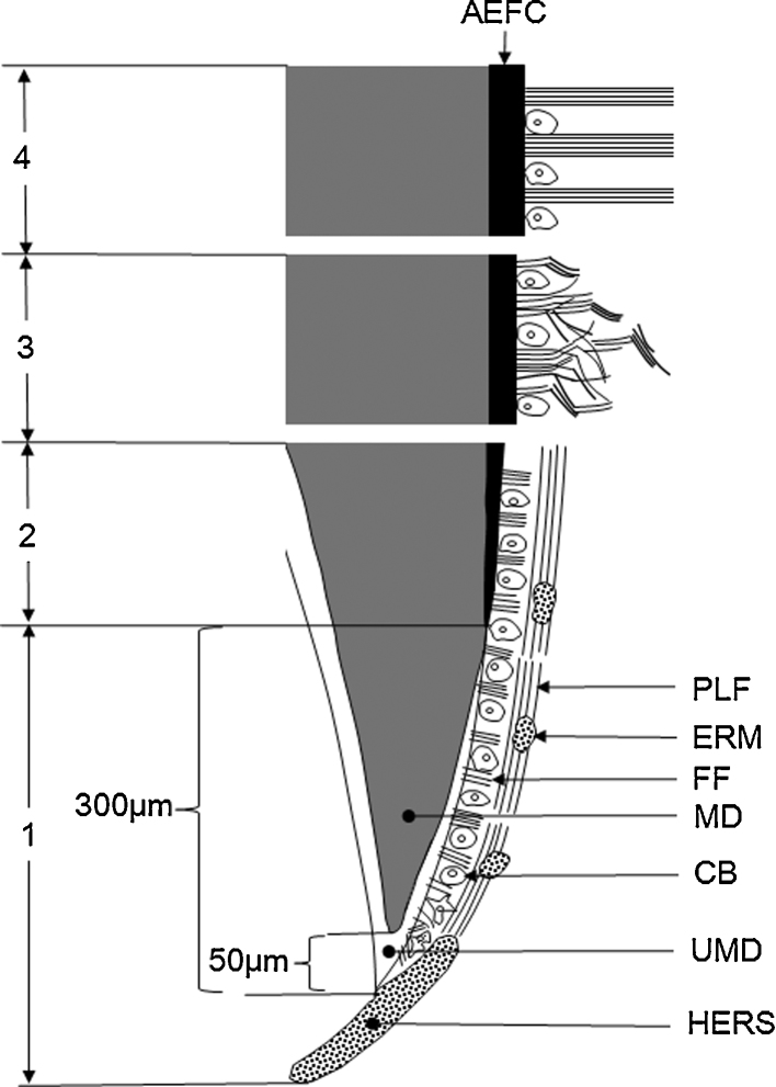
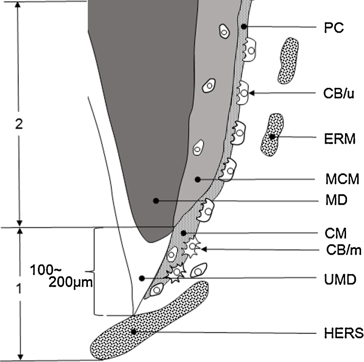
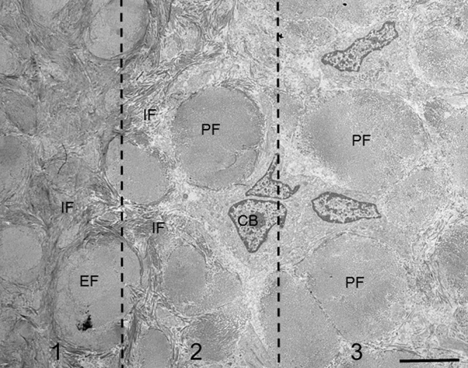
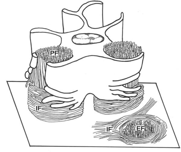
Cementum was first demonstrated by microscopy, about 180 years ago. Since then the biology of cementum has been investigated by the most advanced techniques and equipment at that time in various fields of dental sciences. A great deal of data on cementum histology have been accumulated. These data have been obtained from not only human, but also non-human animals, in particular, rodents such as the mouse and rat. Although many dental histologists have reviewed histology of human cementum, some descriptions are questionable, probably due to incorrect comparison of human and rodent cementum. This review was designed to introduce current histology of human cementum, i.e. its structure, function, and development and to re-examine the most questionable and controversial conclusions made in previous reports.

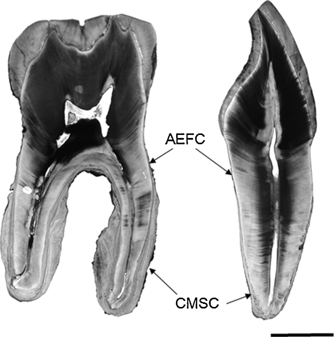
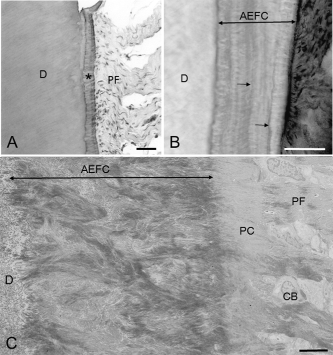
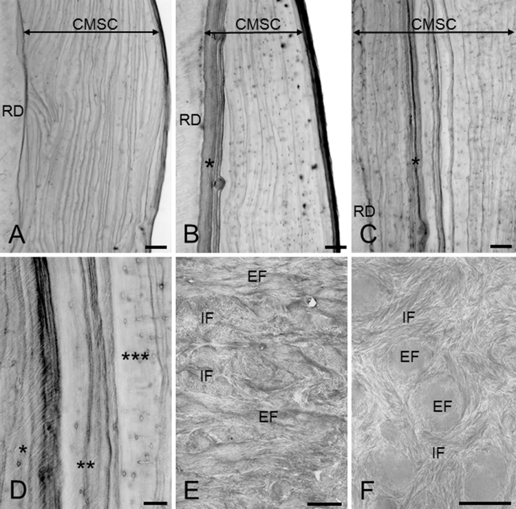
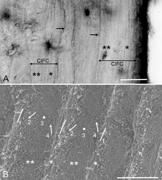
Keywords: Acellular extrinsic fiber cementum; Cellular intrinsic fiber cementum; Cellular mixed stratified cementum; Extrinsic fibers; Human cementum; Intrinsic fibers.


Figures

Figure 1
Figure 1
Full view of a mandibular molar (left) and a maxillary incisor (right). Thin AEFC and thick CMSC cover cervical and apical roots, respectively. CMSC is much thicker in the molar than in the incisor. Hematoxylin-stained ground section. Bar 5 mm.
Figure 2
Figure 2
Light (A, B) and transmission electron (C) micrographs showing AEFC. (A) Principal fibers (PF) enter AEFC (*) as extrinsic fibers. D, dentin. Hematoxylin and eosin-stained paraffin section. Bar 50 μm. (B) Extrinsic fibers are observed as white lines in AEFC. Extrinsic fibers change their orientation at the intensely stainable incremental lines (arrows). D, dentin. Hematoxylin-stained ground section. Bar 50 μm. (C) Extrinsic fibers are branching and anastomosing. D, dentin; CB, cementoblasts; PC, precementum; PF, principal fibers. Bar 5 μm.
Figure 3
Figure 3
(A–D) Micrographs showing structural variety of CMSC in hematoxylin-stained ground sections. CMSC is partitioned by many, intensely stainable incremental lines. RD, root dentin. Bars 100 μm (A–C), 50 μm (D). (A) CMSC consists of stratified CIFC. (B) AEFC (*) is present as the first formed cementum of CMSC. (C) AEFC (*) intervenes between CIFC. (D) Magnification of extrinsic fiber-rich (*), -poor (**), and -free CIFC (***). (E, F) Transmission electron micrographs showing extrinsic (EF) and intrinsic fibers (IF) in CIFC. Bars 5 μm. (E) Extrinsic fibers are branching and anastomosing. Intrinsic fibers fill the space between extrinsic fibers. (F) Intrinsic fibers encircle extrinsic fibers or meander among them in a tangential section parallel to cementum surface.
Figure 4
Figure 4
(A) Magnification of extrinsic fiber-free CIFC partitioned by incremental lines (arrows). In CIFC an alternation of darkly and faintly stainable lamellae is obvious on the periodontal ligament side (*) and non-obvious on the dentin side (**). Bar 30 μm. Hematoxylin-stained ground section. (B) Scanning electron micrograph showing the alternating lamellae. The specimen (a mandibular molar) has been treated by 10% NaOH maceration method to observe individual collagen fibrils clearly. Two types of lamellae, i.e. lamellae of longitudinally and near-longitudinally cut fibril arrays (*) and lamellae of transversely and near-transversely cut fibril arrays (**) create the alternating lamellae. Four types of fibril arrays are roughly recognized: 1, longitudinally cut fibril arrays; 2, obliquely cut fibril arrays facing downward; 3, transversely cut fibril arrays; 4, obliquely cut fibril arrays facing upward. As traced from left to right, the fibril arrays appear to rotate clockwise. Bar 3 μm.
Figure 5
Figure 5
(A–C) Light micrographs showing the apical roots covered with CMSC (A, B) and cervical root covered with AEFC (C) in hematoxylin-stained ground sections. Bars 100 μm. (A, B) The intermediate cementum (*) contains lacunae (small arrows) and exists on the dentin side of the cemento-dentinal junction (large arrow). Arrowheads in (B) indicate the granular layer of Tomes. (C) The hyaline layer of Hopewell-Smith (*) is present between the granular layer of Tomes (arrowheads) and cemento-dentinal junction (arrow).
Figure 6
Figure 6
Schematic diagram depicting AEFC genesis. Section 1: Cementoblasts appear and start to form fiber fringe on the unmineralized dentin. Periodontal ligament fibers are arranged in parallel with the root surface. Section 2: Fiber fringe with maximum density is established. Dentin mineralization reaches the base of fiber fringe and progresses into the fringe. Section 3: Fiber fringe elongates and begins to connect with periodontal ligament fibers. Section 4: The tooth anchorage system, or principal fiber-extrinsic fiber linkage, is established. CB, cementoblasts; ERM, epithelial cell rests of Malassez; FF, fiber fringe; HERS, Hertwig's epithelial root sheath; MD, mineralized dentin; PLF, periodontal ligament fibers; UMD, unmineralized dentin.
Figure 7
Figure 7
(A, B) Cementoblasts on established AEFC in rat molars by transmission (A) and scanning electron microscopy (B). Bars 30 μm. (A) Cementoblasts encircle principal fibers with cytoplasmic processes in a tangential section through cementum surface. (B) The specimen has been treated by KOH-collagenase method and thereby collagen fibers and interfibrillar matrix are removed and only cementoblasts can be selectively observed. Cementoblasts are viewed from the cementum side. Cementoblasts form cylindrical compartments with wing-like processes.
Figure 8
Figure 8
Schematic diagram depicting the formation of extrinsic fiber-free CIFC as the initial CMSC genesis. Section 1: Cementoblasts appear and produce the cementum matrix rapidly in a multipolar mode on the unmineralized dentin. Section 2: Dentin mineralization reaches the cementum matrix and progresses into it. Cementoblasts produce cementum matrix slowly in a unipolar mode. Unmineralized cementum matrix is recognized as precementum. CB/m, cementoblasts with multipolar matrix production; CB/u, cementoblasts with unipolar matrix production; CM, cementum matrix; ERM, epithelial cell rests of Malassez; HERS, Hertwig's epithelial root sheath; MCM, mineralized cementum matrix; MD, mineralized dentin; UMD, unmineralized dentin; PC, precementum.
Figure 9
Figure 9
(A) Transmission electron micrographs showing the CIFC surface where alternating lamellae are generating. Flat cementoblasts cover a lamella (*) of transversely and near-transversely cut fibrils. Bar 2 μm. (B) Magnification of the boxed area in (A). A long, thin process (large arrow) is in a close and parallel association with longitudinally cut fibrils (small arrows). Bar 2 μm.
Figure 10
Figure 10
Transmission electron micrograph showing the extrinsic fiber-rich CIFC surface in a tangential section through the cementum surface. Three sections are divided. Section 1 indicates the interior of cementum, where intrinsic fibers (IF) surround extrinsic fibers (EF). Section 2 indicates the cementum surface. Intrinsic fibers and cementoblasts (CB) surround principal fibers. Section 3 indicates an area slightly distant from the cementum surface. Cementoblasts (CB) surrounds principal fibers with cytoplasmic processes. Only a few or no intrinsic fibers are seen around principal fibers. Bar 5 μm.
Figure 11
Figure 11
Schematic diagram depicting how cementoblasts produce intrinsic fibers around principal fibers on extrinsic fiber-rich CIFC. Cementoblasts surround principal fibers (PF) in cylindrical compartments with wing-like processes. With further cementogenesis, they retract the wing-like processes and divide them into finger-like processes. At the same time they secrete intrinsic fibers (IF) along the finger-like processes. As a result, the intrinsic fibers encircle the extrinsic fibers (EF) in the cementum.
Figure 12
Figure 12
Schematic diagrams depicting the disintegration of Hertwig's epithelial root sheath during root development. (A) Hertwig's epithelial root sheath (HERS) bends inside toward dental pulp (DP) at the tip of developing root (R). (B) Three-dimensionally, the epithelial sheath forms a tapered cylinder. (C) Root formation proceeds almost straight, whereas the epithelial sheath maintains the tapered shape. As a result, due to the discrepancy in surface area, the epithelial sheath is stretched out and fragmented into epithelial cell rests of Malassez.

References

    1. Denton G.B. The discovery of cementum. J Dent Res. 1939;18:239.
    1. Jones S.J. Cement. In: Osborn J.W., editor. Dental anatomy and embryology. Blackwell; Oxford: 1981. pp. 193–205.
    1. Schroeder H.E. Cementum. In: Schroeder H.E., editor. The periodontium. Springer; Berlin: 1986. pp. 23–127.
    1. Schroeder H.E. Biological problems of regenerative cementogenesis: synthesis and attachment of collagenous matrices on growing and established root surface. Int Rev Cytol. 1992;142:1–59. - PubMed
    1. Schroeder H.E. Human cellular mixed stratified cementum: a tissue with alternating layers of acellular extrinsic- and cellular intrinsic fiber cementum. Schweiz Monatsschr Zahnmed. 1993;103:550–560. - PubMed

LinkOut - more resources