Long-Term Effects of Maternal Deprivation on Redox Regulation in Rat Brain: Involvement of NADPH Oxidase
- PMID: 28408971
- PMCID: PMC5376945
- DOI: 10.1155/2017/7390516
Long-Term Effects of Maternal Deprivation on Redox Regulation in Rat Brain: Involvement of NADPH Oxidase
Abstract
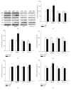
Maternal deprivation (MD) causes perinatal stress, with subsequent behavioral changes which resemble the symptoms of schizophrenia. The NADPH oxidase is one of the major generators of reactive oxygen species, known to play a role in stress response in different tissues. The aim of this study was to elucidate the long-term effects of MD on the expression of NADPH oxidase subunits (gp91phox, p22phox, p67phox, p47phox, and p40phox). Activities of cytochrome C oxidase and respiratory chain Complex I, as well as the oxidative stress parameters using appropriate spectrophotometric techniques were analyzed. Nine-day-old Wistar rats were exposed to a 24 h maternal deprivation and sacrificed at young adult age. The structures affected by perinatal stress, cortex, hippocampus, thalamus, and caudate nuclei were investigated. The most prominent findings were increased expressions of gp91phox in the cortex and hippocampus, increased expression of p22phox and p40phox, and decreased expression of gp91phox, p22phox, and p47phox in the caudate nuclei. Complex I activity was increased in all structures except cortex. Content of reduced glutathione was decreased in all sections while region-specific changes of other oxidative stress parameters were found. Our results indicate the presence of long-term redox alterations in MD rats.
Figures


Similar articles
-
Intracellular localization and preassembly of the NADPH oxidase complex in cultured endothelial cells.J Biol Chem. 2002 May 31;277(22):19952-60. doi: 10.1074/jbc.M110073200. Epub 2002 Mar 13. J Biol Chem. 2002. PMID: 11893732
-
p40(phox) Participates in the activation of NADPH oxidase by increasing the affinity of p47(phox) for flavocytochrome b(558).Biochem J. 2000 Jul 1;349(Pt 1):113-7. doi: 10.1042/0264-6021:3490113. Biochem J. 2000. PMID: 10861218 Free PMC article.
-
Molecular characterization and localization of the NAD(P)H oxidase components gp91-phox and p22-phox in endothelial cells.Arterioscler Thromb Vasc Biol. 2000 Aug;20(8):1903-11. doi: 10.1161/01.atv.20.8.1903. Arterioscler Thromb Vasc Biol. 2000. PMID: 10938010
-
NADPH oxidase activation in neutrophils: Role of the phosphorylation of its subunits.Eur J Clin Invest. 2018 Nov;48 Suppl 2:e12951. doi: 10.1111/eci.12951. Epub 2018 Jun 3. Eur J Clin Invest. 2018. PMID: 29757466 Review.
-
Interactions between the components of the human NADPH oxidase: intrigues in the phox family.J Lab Clin Med. 1996 Nov;128(5):461-76. doi: 10.1016/s0022-2143(96)90043-8. J Lab Clin Med. 1996. PMID: 8900289 Review.
Cited by
-
Animal Model of Autism Induced by Valproic Acid Combined with Maternal Deprivation: Sex-Specific Effects on Inflammation and Oxidative Stress.Mol Neurobiol. 2025 Mar;62(3):3653-3672. doi: 10.1007/s12035-024-04491-z. Epub 2024 Sep 24. Mol Neurobiol. 2025. PMID: 39316355
-
The long-term effects of maternal deprivation on the number and size of inhibitory interneurons in the rat amygdala and nucleus accumbens.Front Neurosci. 2023 Jun 26;17:1187758. doi: 10.3389/fnins.2023.1187758. eCollection 2023. Front Neurosci. 2023. PMID: 37434764 Free PMC article.
-
Maternal Deprivation in Rats Decreases the Expression of Interneuron Markers in the Neocortex and Hippocampus.Front Neuroanat. 2021 Jun 8;15:670766. doi: 10.3389/fnana.2021.670766. eCollection 2021. Front Neuroanat. 2021. PMID: 34168541 Free PMC article.
-
Environmental Enrichment Rescues Oxidative Stress and Behavioral Impairments Induced by Maternal Care Deprivation: Sex- and Developmental-Dependent Differences.Mol Neurobiol. 2023 Dec;60(12):6757-6773. doi: 10.1007/s12035-021-02588-3. Epub 2021 Oct 19. Mol Neurobiol. 2023. PMID: 34665408
-
Proteome profiling of different rat brain regions reveals the modulatory effect of prolonged maternal separation on proteins involved in cell death-related processes.Biol Res. 2021 Feb 8;54(1):4. doi: 10.1186/s40659-021-00327-5. Biol Res. 2021. PMID: 33557947 Free PMC article.
References
-
- Viveros M. P., Diaz F., Mateos B., Rodriguez N., Chowen J. A. Maternal deprivation induces a rapid decline in circulating leptin levels and sexually dimorphic modifications in hypothalamic trophic factors and cell turnover. Hormones and Behavior. 2010;58(5):808–819. doi: 10.1016/j.yhbeh.2010.08.003. - DOI - PubMed
-
- Ellenbroek B. A., Cools A. R. Animal models with construct validity for schizophrenia. Behavioural Pharmacology. 1990;1(6):469–490. - PubMed
MeSH terms
Substances
LinkOut - more resources
Full Text Sources
Other Literature Sources

