. 2017 Feb 20;6(1):e219-e225.
doi: 10.1016/j.eats.2016.09.025. eCollection 2017 Feb.

Multidirectional Shoulder Instability: Arthroscopic Labral Augmentation

Affiliations

Multidirectional Shoulder Instability: Arthroscopic Labral Augmentation

Enrico Gervasi et al. Arthrosc Tech. .

Abstract

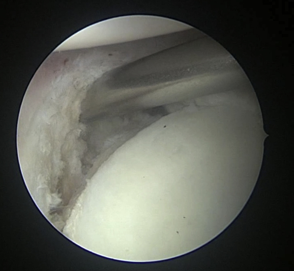
Capsulolabral augmentation is one of the most used arthroscopic techniques to address multidirectional instability of the shoulder. Given the thin and weak capsule seen in the affected patients, reconstruction in this subset of patients can be particularly challenging. This arthroscopic technique aims to reduce the capsular volume and deepen the glenoid socket through the creation of a particularly voluminous "bumper" along the glenoid bone. Increasing the depth of the glenoid facilitates a concavity-compression stabilizing effect and, therefore, shoulder stability, especially midrange stability. This technique aims to augment the bump of the standard capsulolabral reconstruction by using a resorbable surgical mesh derived from porcine skin.


Figures

Fig 1
Fig 1
Patient in lateral decubitus, left shoulder. The 3 portals held by a cannula are visible: posterior, anterosuperior, and midglenoid. The anterosuperior portal is mainly used for intra-articular viewing during the procedure.
Fig 2
Fig 2
Patient in lateral decubitus, left shoulder, posterior viewing of the left shoulder. A posterolateral portal (dermographic pen) can be used for a better anchor placement in the inferior portion of the glenoid bone (6 o'clock). A red arrow marks the standard posterior portal.
Fig 3
Fig 3
Patient in lateral decubitus, left shoulder, camera in the anterosuperior portal. Capsuloligamentous structures already detached from the subequatorial portion of the glenoid bone. The tool lifts the disconnected medial margin of the capsule and shows the decorticated glenoid neck.
Fig 4
Fig 4
Patient in lateral decubitus, left shoulder, camera in the anterosuperior portal. Capsulolabroplasty is performed using double-loaded all-suture anchors. Anchor threads at the 6 o'clock position already passed through the capsular tissue; using a shuttle technique, we are working in the posteroinferior quadrant, with the second anchor seated at the 4.30 position. In this phase, a South-North shift of the capsular tissue needs to be obtained; therefore, the tool must emerge distally from the anchor.
Fig 5
Fig 5
Patient in lateral decubitus, left shoulder, camera in the anterosuperior portal. The capsulolabroplasty is finished, the capsuloligamentous structures are retensioned, and a new glenoid labrum (called “bumper”) is reconstructed.
Fig 6
Fig 6
The membrane is folded to assume a cylindrical shape. Two surgical clamps fix the membrane at the extremities. To keep the shape during suturing and ease the needle passage, straight 18-gauge needles pass through the folded graft. All needles have to be inserted in the same direction.
Fig 7
Fig 7
The Vicryl No.1 (Ethicon, Somerville, NJ), tied to form a loop, is inserted in an eyed needle. Passing through the straight needles, the eyed needle pierces the graft and the suture thread along with it. The same procedure is to be performed with further needles, managing the suture with a SpeedWhip technique.
Fig 8
Fig 8
A resorbable thread keeps the cylindrical shape of the graft. Three LabralTapes (Arhtrex, Naples, FL) pass through the graft at both ends and in the center. The free limbs of each LabralTape come out of the same side of the graft to shape a “U.” The central LabralTape has a different color to ease suture management within the articulation. We recommend tying together the loose ends of each LabralTape. The 2 LabralTapes at the extremities are used to anchor the graft to the anterior and to the posterior margins of the glenoid. The central LabralTape is used to anchor the graft at the 6 o'clock position. The posterior LabralTape is used to pull the graft inside the joint.
Fig 9
Fig 9
One of the LabralTapes at the extremity of the graft is inserted into the articulation through the midglenoid portal and retrieved from the posterior one. Applying traction to this LabralTape, the graft is pulled into the joint through the midglenoid portal.
Fig 10
Fig 10
Patient in lateral decubitus, left shoulder, camera in the anteroposterior portal. Once a hole has been made at the 6 o'clock position, the central LabralTape (the one differently colored) is anchored to the glenoid with a knotless anchor through the midglenoid portal.
Fig 11
Fig 11
Patient in lateral decubitus, left shoulder, camera in the anteroposterior portal. The graft is anchored to the posterior margin of the glenoid with a knotless anchor (red arrow) through the posterior portal.
Fig 12
Fig 12
Patient in lateral decubitus, left shoulder, camera in the anteroposterior portal. The graft made of resorbable surgical mesh derived from porcine skin is anchored to the glenoid rim along the entire subequatorial region.

References

    1. Gaskill T.R., Taylor D.C., Millet P.J. Management of multidirectional instability of the shoulder. J Am Acad Orthop Surg. 2011;19:758–767. - PubMed
    1. Metcalf M.H., Pon D.J., Harryman D.T., II Capsulolabral augmentation increase glenohumeral stability in the cadaver shoulder. J Shoulder Elbow Surg. 2001;10:532–538. - PubMed
    1. Apostolakos J., Yang J.S., Hoberman A.R. Glenoid labrum. In: Bain G.I., Itoi E., Di Giacomo G., Sugaya H., editors. Normal and pathological anatomy of the shoulder. Springer-Verlag; Berlin: 2015. pp. 83–91.
    1. Warner J.J., Bowen M.K., Deng X., Torzilli P.A., Warren R.F. Effect of joint compression on inferior stability of the glenohumeral joint. J Shoulder Elbow Surg. 1999;11:141–149. - PubMed
    1. Lippitt S.B., Vanderhooft J.E., Harris S.L., Sidles J.A., Harryman D.T., Matsen F.A. Glenohumeral stability from concavity-compression: A quantitative analysis. J Shoulder Elbow Surg. 1993;2:27–35. - PubMed

LinkOut - more resources