

Axonal damage and loss of connectivity in nigrostriatal and mesolimbic dopamine pathways in early Parkinson's disease
- PMID: 28409113
- PMCID: PMC5379906
- DOI: 10.1016/j.nicl.2017.03.011
Axonal damage and loss of connectivity in nigrostriatal and mesolimbic dopamine pathways in early Parkinson's disease
Abstract
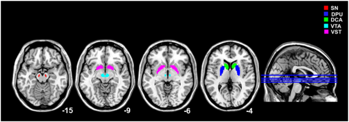
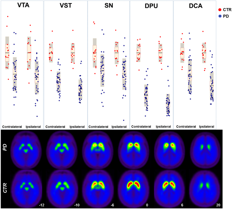
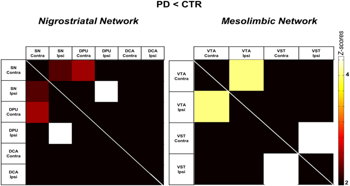
A progressive loss of dopamine neurons in the substantia nigra (SN) is considered the main feature of idiopathic Parkinson's disease (PD). Recent neuropathological evidence however suggests that the axons of the nigrostriatal dopaminergic system are the earliest target of α-synuclein accumulation in PD, thus the principal site for vulnerability. Whether this applies to in vivo PD, and also to the mesolimbic system has not been investigated yet. We used [11C]FeCIT PET to measure presynaptic dopamine transporter (DAT) activity in both nigrostriatal and mesolimbic systems, in 36 early PD patients (mean disease duration in months ± SD 21.8 ± 10.7) and 14 healthy controls similar for age. We also performed anatomically-driven partial correlation analysis to evaluate possible changes in the connectivity within both the dopamine networks at an early clinical phase. In the nigrostriatal system, we found a severe DAT reduction in the afferents to the dorsal putamen (DPU) (η2 = 0.84), whereas the SN was the less affected region (η2 = 0.31). DAT activity in the ventral tegmental area (VTA) and the ventral striatum (VST) were also reduced in the patient group, but to a lesser degree (VST η2 = 0.71 and VTA η2 = 0.31). In the PD patients compared to the controls, there was a marked decrease in dopamine network connectivity between SN and DPU nodes, supporting the significant derangement in the nigrostriatal pathway. These results suggest that neurodegeneration in the dopamine pathways is initially more prominent in the afferent axons and more severe in the nigrostriatal system. Considering PD as a disconnection syndrome starting from the axons, it would justify neuroprotective interventions even if patients have already manifested clinical symptoms.
Keywords: AI, asymmetry index; Axonal damage; DCA, dorsal caudate; DPU, dorsal putamen; Dopamine transporter; Molecular connectivity; Parkinson's disease; Positron emission tomography; SN, substantia nigra; SUVr, standardized uptake value ratio; VST, ventral striatum; VTA, ventral tegmental area; cAS, clinical asymmetry.
Figures
References
-
- Ba F., Martin W.R.W. Dopamine transporter imaging as a diagnostic tool for parkinsonism and related disorders in clinical practice. Parkinsonism Relat. Disord. 2015;21:87–94. - PubMed
-
- Braak H., Ghebremedhin E., Rüb U., Bratzke H., Del Tredici K. Stages in the development of Parkinson's disease-related pathology. Cell Tissue Res. 2004;318:121–134. - PubMed
-
- Calo L., Wegrzynowicz M., Santivañez-Perez J., Grazia Spillantini M. Synaptic failure and α-synuclein. Mov. Disord. 2016;0 (n/a-n/a) - PubMed
Publication types
MeSH terms
Substances
LinkOut - more resources
Full Text Sources
Other Literature Sources
