

The Association between Imaging Parameters of the Paraspinal Muscles, Spinal Degeneration, and Low Back Pain
- PMID: 28409152
- PMCID: PMC5376928
- DOI: 10.1155/2017/2562957
The Association between Imaging Parameters of the Paraspinal Muscles, Spinal Degeneration, and Low Back Pain
Abstract
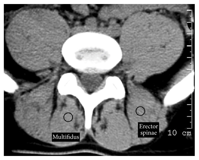
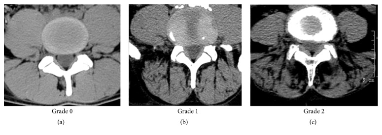
This narrative review investigated imaging parameters of the paraspinal muscles and their association with spinal degenerative features and low back pain (LBP) found in the literature. Three principal signs of muscle degeneration were detected on imaging: decreased muscle size, decreased radiographic density, and increased fat deposits. Men have a higher density of paraspinal muscles than women, younger individuals have a higher density than older ones, and lean individuals have a higher density than those with an increased body mass index. Fatty infiltration appears to be a late stage of muscular degeneration and can be measured noninvasively by an MRI scan. Fatty infiltration in the lumbar multifidus is common in adults and is strongly associated with LBP, especially in women, independent of body composition. Fatty infiltration develops in areas where most degenerative changes are found. MR spectroscopy studies have corroborated that the lumbar multifidus in LBP subjects has a significantly higher fat content than asymptomatic controls. There is a strong need for establishing uniform methods of evaluating normal parameters and degenerative changes of the paraspinal muscles. Additional imaging studies are needed to improve the understanding of the association and causal relationships between LBP, spinal degeneration, and changes in the paraspinal muscles.
Figures
References
Publication types
MeSH terms
LinkOut - more resources
Full Text Sources
Other Literature Sources
Miscellaneous
