Review
. 2017 Jun;133(6):863-885.
doi: 10.1007/s00401-017-1708-8. Epub 2017 Apr 13.

Motor neuron vulnerability and resistance in amyotrophic lateral sclerosis

Affiliations
Review

Motor neuron vulnerability and resistance in amyotrophic lateral sclerosis

Jik Nijssen et al. Acta Neuropathol. 2017 Jun.

Abstract

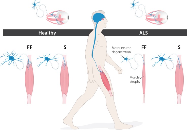
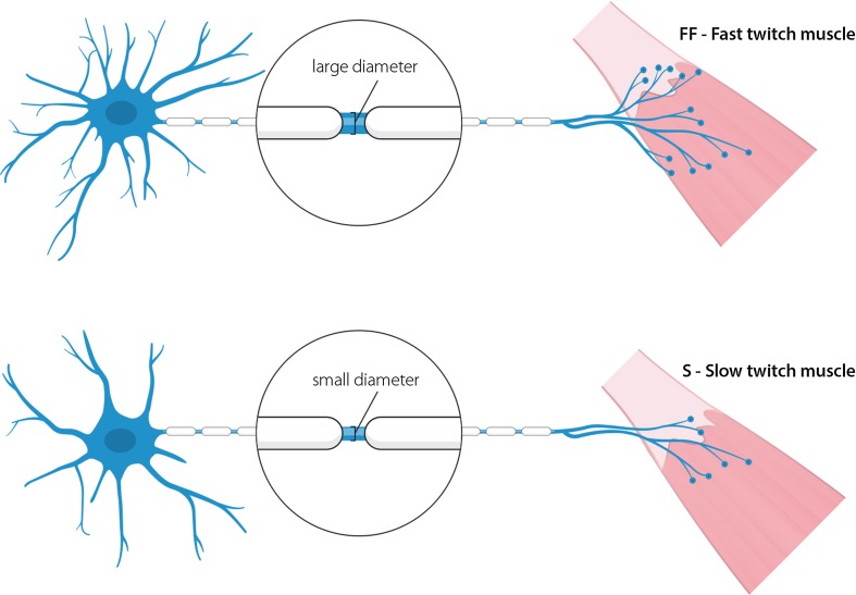
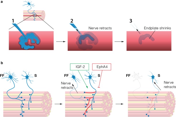
In the fatal disease-amyotrophic lateral sclerosis (ALS)-upper (corticospinal) motor neurons (MNs) and lower somatic MNs, which innervate voluntary muscles, degenerate. Importantly, certain lower MN subgroups are relatively resistant to degeneration, even though pathogenic proteins are typically ubiquitously expressed. Ocular MNs (OMNs), including the oculomotor, trochlear and abducens nuclei (CNIII, IV and VI), which regulate eye movement, persist throughout the disease. Consequently, eye-tracking devices are used to enable paralysed ALS patients (who can no longer speak) to communicate. Additionally, there is a gradient of vulnerability among spinal MNs. Those innervating fast-twitch muscle are most severely affected and degenerate first. MNs innervating slow-twitch muscle can compensate temporarily for the loss of their neighbours by re-innervating denervated muscle until later in disease these too degenerate. The resistant OMNs and the associated extraocular muscles (EOMs) are anatomically and functionally very different from other motor units. The EOMs have a unique set of myosin heavy chains, placing them outside the classical characterization spectrum of all skeletal muscle. Moreover, EOMs have multiple neuromuscular innervation sites per single myofibre. Spinal fast and slow motor units show differences in their dendritic arborisations and the number of myofibres they innervate. These motor units also differ in their functionality and excitability. Identifying the molecular basis of cell-intrinsic pathways that are differentially activated between resistant and vulnerable MNs could reveal mechanisms of selective neuronal resistance, degeneration and regeneration and lead to therapies preventing progressive MN loss in ALS. Illustrating this, overexpression of OMN-enriched genes in spinal MNs, as well as suppression of fast spinal MN-enriched genes can increase the lifespan of ALS mice. Here, we discuss the pattern of lower MN degeneration in ALS and review the current literature on OMN resistance in ALS and differential spinal MN vulnerability. We also reflect upon the non-cell autonomous components that are involved in lower MN degeneration in ALS.

Keywords: ALS; Fast and slow motor units; Neurodegeneration; Neuromuscular junction; Oculomotor neuron; Selective vulnerability.


Figures

Fig. 1
Fig. 1
Levels of selective vulnerability in ALS. ALS selectively affects the somatic motor neuron system. Specific motor neuron pools are more resistant, such as the oculomotor neurons, which persist until end-stage of disease. Additionally, subtypes of affected spinal motor neurons also display a gradient of vulnerability. Fast-twitch fast-fatigable (FF) motor neurons degenerate before slow (S) motor neurons
Fig. 2
Fig. 2
Neuromuscular endplates in extraocular muscle. All six extraocular muscles in mammals have a unique arrangement of neuromuscular endplates. Two distinct bands (rather than one) can be observed (a). Extraocular muscles contain a unique band of small en grappe endplates (b, d), besides the regular band of normal-sized en plaque endplates (c, e). Multiple innervation occurs between bands, such that one muscle fibre can have multiple NMJs, one within each endplate band. Scale bars 50 μm, bars in c and e also apply also to b and d, respectively. AchR acetylcholine-receptor, SV2/NF synaptic vesicle protein 2 and neurofilament 165 kDa
Fig. 3
Fig. 3
Comparison of a spinal motor unit and an oculomotor unit. Ocular motor units differ from spinal motor units in several key aspects. Oculomotor neurons have smaller somas and their dendritic tree is not as complex as that of a spinal motor neuron. Oculomotor neurons innervate multiple muscle fibre types, whereas spinal motor neurons are generally restricted to the innervation of a single type of muscle fibre. Extraocular muscle itself is more complex, containing many different fibre types, not only those containing the classical skeletal myosins. Oculomotor neurons innervate very few endplates compared to spinal motor neurons, but poly-innervation occurs on a large scale
Fig. 4
Fig. 4
Subtypes of spinal motor neurons. FF (fast-twitch, fast-fatigable) and S (slow-twitch) are the two extremes of spinal motor neurons. FF motor neurons have bigger and more complex dendritic trees and project with larger calibre axons. FF motor neurons innervate a large number of endplates and are thus more involved in high-force movements. S motor neurons are more finely structured and are more suited to their function in slow movements and posture maintenance
Fig. 5
Fig. 5
Motor neuron degeneration in ALS. a A dying motor neuron retracts its axon from the neuromuscular junction. The axon continues to die back towards the soma. Meanwhile the endplate on the muscle fibre dissolves and shrinks as acetylcholine receptors are internalised and removed. b FF motor neurons are the most vulnerable among spinal motors. S motor neurons can temporarily compensate for the loss of FF motor neurons by axonal sprouting and re-innervation of denervated endplates. Overexpression of EphA4 can reduce this effect, while overexpression of IGF-2 stimulates re-innervation, causing ALS mice to live longer. Ultimately, however, also S motor neurons die back and leave the muscle denervated
None
None

References

    1. Abhinav K, Stanton B, Johnston C, Hardstaff J, Orrell R, Howard R, Clarke J, Sakel M, Ampong M, Shaw C. Amyotrophic lateral sclerosis in South-East England: a population-based study. Neuroepidemiology. 2007;29:44–48. doi: 10.1159/000108917. - DOI - PubMed
    1. Al-Chalabi A, Andersen PM, Chioza B, Shaw C, Sham PC, Robberecht W, Matthijs G, Camu W, Marklund SL, Forsgren L. Recessive amyotrophic lateral sclerosis families with the D90A SOD1 mutation share a common founder: evidence for a linked protective factor. Hum Mol Genet. 1998;7:2045–2050. doi: 10.1093/hmg/7.13.2045. - DOI - PubMed
    1. Alexianu ME, Ho BK, Mohamed AH, La Bella V, Smith RG, Appel SH. The role of calcium-binding proteins in selective motoneuron vulnerability in amyotrophic lateral sclerosis. Ann Neurol. 1994;36:846–858. doi: 10.1002/ana.410360608. - DOI - PubMed
    1. Allodi I, Comley L, Nichterwitz S, Nizzardo M, Simone C, Benitez JA, Cao M, Corti S, Hedlund E. Differential neuronal vulnerability identifies IGF-2 as a protective factor in ALS. Sci Rep. 2016;6:25960. doi: 10.1038/srep25960. - DOI - PMC - PubMed
    1. An T, Shi P, Duan W, Zhang S, Yuan P, Li Z, Wu D, Xu Z, Li C, Guo Y. Oxidative stress and autophagic alteration in brainstem of SOD1-G93A mouse model of ALS. Mol Neurobiol. 2014;49:1435–1448. doi: 10.1007/s12035-013-8623-3. - DOI - PubMed

Publication types

MeSH terms