. 2017 Apr 14;58(2):118-127.
doi: 10.3325/cmj.2017.58.118.

Antiplatelet and anti-proliferative action of disintegrin from Echis multisquamatis snake venom

Affiliations

Antiplatelet and anti-proliferative action of disintegrin from Echis multisquamatis snake venom

Volodymyr Chernyshenko et al. Croat Med J. .

Abstract

Aim: To purify the platelet aggregation inhibitor from Echis multisquamatis snake venom (PAIEM) and characterize its effect on platelet aggregation and HeLa cell proliferation.

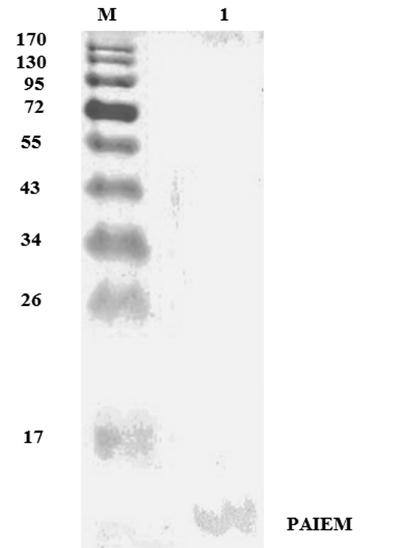
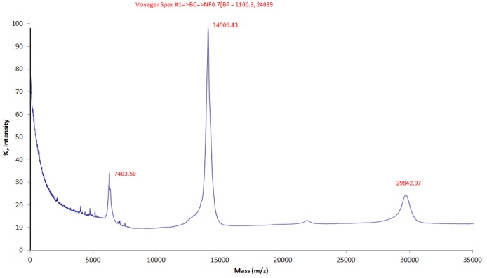
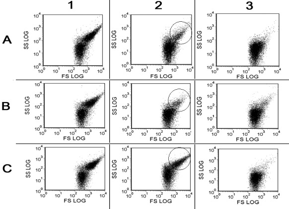
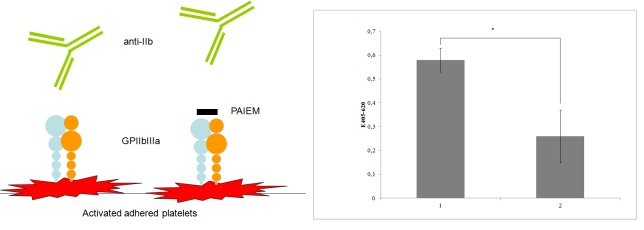
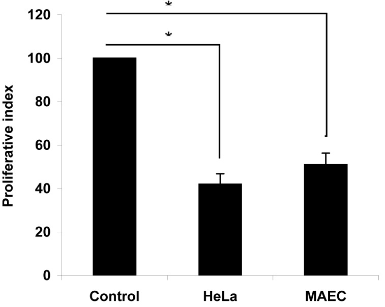
Methods: Sodium dodecyl sulfate-polyacrylamide gel electrophoresis (SDS-PAGE) and matrix assisted laser desorption/ionization time-of-flight (MALDI-TOF) were used for PAIEM identification. Platelet aggregation in the presence of PAIEM was studied on aggregometer Solar-AP2110. The changes of shape and granularity of platelets in the presence of PAIEM were studied on flow cytometer COULTER EPICS XL, and degranulation of platelets was estimated using spectrofluorimetry. Indirect enzyme-linked immunosorbent assay was used for the determination of target of PAIEM on platelet surface. An assay based on 3-(4,5-dimethylthiazol-2-yl)-2,5-diphenyltetrazolium bromide was used to evaluate the effect of PAIEM on the proliferation of HeLa cells in cell culture.

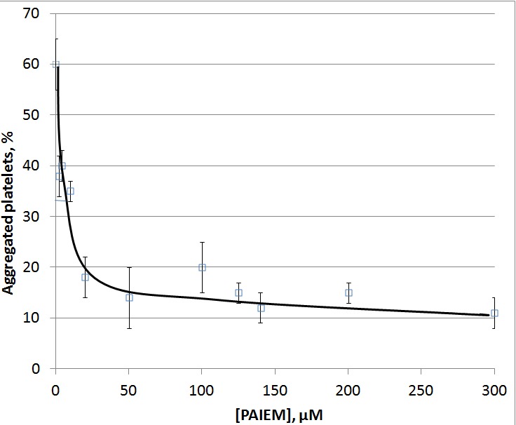
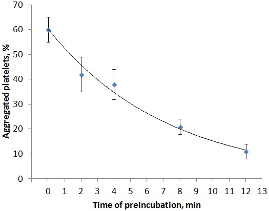
Results: The molecular weight of the protein purified from Echis multisquamatis venom was 14.9 kDa. Half-maximal inhibitory concentration (IC50) of PAIEM needed to inhibit adenosine diphosphate (ADP)-induced platelet aggregation was 7 μM. PAIEM did not affect thrombin- or ADP-induced platelet activation, but it did prevent binding of the anti-IIb antibody to glycoprotein IIb/IIIa (GPIIbIIIa)-receptor of adhered platelets and inhibited the viability of HeLa cells by 54%.

Conclusion: As a member of the disintegrin family, PAIEM inhibited platelet aggregation and cell proliferation possibly by blocking integrin-mediated interactions. However, it did not impair cellular signaling causing any changes in platelet shape and granularity and did not affect ADP-induced platelet degranulation. This disintegrin was shown to be a potent inhibitor of integrin-mediated cellular interactions including platelet aggregation or cancer cell proliferation.


Figures

Figure 1
Figure 1
Sodium dodecyl sulfate-polyacrylamide gel electrophoresis (SDS-PAGE) of platelet aggregation inhibitor from the venom of Echis multisquamatis (PAIEM) eluted from Q-Sepharose and then purified on Superdex G-75 column. M – molecular weight markers.
Figure 2
Figure 2
Matrix Assisted Laser Desorption/Ionization Time-of-Flight (MALDI-TOF) analysis of purified platelet aggregation inhibitor from the Echis multisquamatis (PAIEM) snake venom. According to MALDI-TOF spectrometry, the protein has molecular weight 14.9 kDa. Other two peaks marked on the graph correspond to 1/2 and 2 charged molecules.
Figure 3
Figure 3
Dose-dependence curve of the level of adenosine diphosphate-induced (2.5 µM) aggregation of human platelets in platelet rich plasma in the presence of platelet aggregation inhibitor from the Echis multisquamatis (PAIEM) snake venom. Boxes and whiskers represent the mean values ± standard error, respectively (data of 3 typical experiments made in triplicate). *P < 0.05 as compared to the control.
Figure 4
Figure 4
The effect of preincubation of platelet-rich plasma with 5 μg/mL of platelet aggregation inhibitor from the Echis multisquamatis (PAIEM) snake venom for 2, 4, 6, 8 12 minutes on the aggregation of platelets induced by 2.5 μM of adenosine diphospate. Data represent the mean values ± standard error (data of 3 typical experiments made in triplicate), *P < 0.05 as compared to the control.
Figure 5
Figure 5
Flow cytometry of washed human platelets activated by 0.125 NIH/mL of thrombin. 1-3 – 1, 2, and 3 minutes after the stimulation, respectively. A. – control probe; B. – in the presence of 50 μg/mL of platelet aggregation inhibitor from the Echis multisquamatis (PAIEM) snake venom; C. – in the presence of 1% dimethyl sulfoxide as the inhibitor of platelet activation. SS – side light scattering, parameter of platelets granulation: FS – frontal light scattering, parameter of platelets shape. Traces are typical for 3 independent experiments made in triplicate.
Figure 6
Figure 6
The acidification of platelets secretory granules and the release of granule constituents during adenosine diphosphate (ADP)-induced activation in the presence of platelet aggregation inhibitor from the Echis multisquamatis (PAIEM) snake venom. Platelets were loaded with pH-sensitive fluorescent dye acridine orange and stimulated with 2 μM of ADP in the presence or absence of PAIEM. Traces are typical for 5 independent experiments. A – after the addition of equivalent amount of buffer; B – in the presence of 10 μg/mL of PAIEM; C – in the presence of 1% dimethyl sulfoxide; D – summarized enlarged fragment of graphics A-C. Traces are typical for 3 independent experiments performed 5 times each.
Figure 7
Figure 7
A. Scheme of experiment that confirmed the platelet aggregation inhibitor from the Echis multisquamatis (PAIEM) snake venom binding to glycoprotein (GP)IIb/IIIa. Washed human platelets were added to the wells of 96-well tissue culture plate and then activated by 30 µM of adenosine diphosphate. Antibody CD61 (specific to IIb subunit of Iib/IIIa platelet receptor) was added to the first row of wells, antibody CD61 was added to the second row alongside with PAIEM (0.05 mg/mL). B. Enzyme-linked immunosorbent assay of anti-GPIIb binding to activated platelets in the presence of PAIEM. Boxes and whiskers represent the mean values ± standard error (data of 3 typical experiments performed 5 times each), *P < 0.05 as compared to the control.
Figure 8
Figure 8
Proliferative index of HeLa and mouse aortic endothelial cells (MAEC) cells in the presence of 6 µg/mL of the platelet aggregation inhibitor from the Echis multisquamatis (PAIEM) snake venom vs equivalent volume of tris-buffered saline (Control). Cell proliferation rate was calculated as [(1−ODexperimental group)/ODcontrol group] × 100%. The absorbance was measured at 540 nm. Boxes and whiskers represent the mean values ± standard error, *P < 0.05 as compared to the control.

Similar articles

Cited by

References

    1. Jackson SP. The growing complexity of platelet aggregation. Blood. 2007;109:5087–95. doi: 10.1182/blood-2006-12-027698. - DOI - PubMed
    1. Offermanns S. Activation of platelet function through G protein-coupled receptors. Circ Res. 2006;99:1293–304. doi: 10.1161/01.RES.0000251742.71301.16. - DOI - PubMed
    1. Wijeyeratne YD, Heptinstall S. Anti-platelet therapy: ADP receptor antagonists. Br J Clin Pharmacol. 2011;72:647–57. doi: 10.1111/j.1365-2125.2011.03999.x. - DOI - PMC - PubMed
    1. Dorsam RT, Kunapuli SP. Central role of the P2Y12 receptor in platelet activation. J Clin Invest. 2004;113:340–5. doi: 10.1172/JCI20986. - DOI - PMC - PubMed
    1. Schrör K. Aspirin and platelets: the antiplatelet action of aspirin and its role in thrombosis treatment and prophylaxis. Semin Thromb Hemost. 1997;23:349–56. doi: 10.1055/s-2007-996108. - DOI - PubMed

Substances