. 2017 Jul;174(13):2085-2101.
doi: 10.1111/bph.13820. Epub 2017 May 27.

A sphingosine-1-phosphate receptor type 1 agonist, ASP4058, suppresses intracranial aneurysm through promoting endothelial integrity and blocking macrophage transmigration

Affiliations

A sphingosine-1-phosphate receptor type 1 agonist, ASP4058, suppresses intracranial aneurysm through promoting endothelial integrity and blocking macrophage transmigration

Rie Yamamoto et al. Br J Pharmacol. 2017 Jul.

Abstract

Background and purpose: Intracranial aneurysm (IA), common in the general public, causes lethal subarachnoid haemorrhage on rupture. It is, therefore, of utmost importance to prevent the IA from rupturing. However, there is currently no medical treatment. Recent studies suggest that IA is the result of chronic inflammation in the arterial wall caused by endothelial dysfunction and infiltrating macrophages. The sphingosine-1-phosphate receptor type 1 (S1P1 receptor) is present on the endothelium and promotes its barrier function. Here we have tested the potential of an S1P1 agonist, ASP4058, to prevent IA in an animal model.

Experimental approach: The effects of a selective S1P1 agonist, ASP4058, on endothelial permeability and migration of macrophages across an endothelial cell monolayer were tested in vitro using a Transwell system, and its effects on the size of IAs were evaluated in a rat model of IA.

Key results: S1P1 receptor was expressed in endothelial cells of human IA lesions and control arterial walls. ASP4058 significantly reduced FITC-dextran leakage through an endothelial monolayer and suppressed the migration of macrophages across the monolayer in vitro. Oral administration of ASP4058 reduced the vascular permeability, macrophage infiltration and size of the IAs by acting as an S1P1 agonist in the rat model. This effect was mimicked by another two structurally-unrelated S1P1 agonists.

Conclusion and implications: A selective S1P1 agonist is a strong drug candidate for IA treatment as it promotes the endothelial cell barrier and suppresses the trans-endothelial migration of macrophages in IA lesions.


Figures

Figure 1
Figure 1
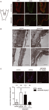
Expression of S1P1 receptors in human control arterial wall (A) and IA lesion (B, C). Adjacent sections were prepared from branches of carotid artery or IA lesion of humans and immunostained for α‐ SMA, a medial smooth muscle marker, CD31, an endothelial cell marker and S1P1 receptors. Merged images for CD31 and S1P1 staining, SMA and S1P1 staining and images with Haematoxylin–Eosin staining (HE) are also shown. Box in the left panels indicates the region magnified in the following panels. Bar, 50 μm.
Figure 2
Figure 2
Inhibition of endothelial permeability by ASP4058 mediated by the S1P1 receptor. (A) Expression of each S1P receptor subtype in primary culture of endothelial cells (HCtAECs). Primer set to detect β‐actin mRNA served as a positive control (P). (B) Concentration‐dependent inhibition of forskolin‐induced cAMP accumulation in HCtAECs by ASP4058. HCtAECs were treated with 1 μM forskolin in the presence of the indicated concentrations of ASP4058, and cAMP accumulation was examined as described in the Methods. Data represent mean ± SEM (n = 5). * P < 0.05. (C, D) Effect of ASP4058 on surface expression of S1P1 receptors in HCtAECs. HCtAECs were treated with the indicated concentrations of ASP4058 for 1 h, and the surface expression of S1P1 receptors was examined by FACS. Representative histogram of FACS analysis for S1P1 receptor is shown in (C) (n = 5). (D) The surface expression of S1P1 receptors calculated as described in the Methods. Data represents mean ± SEM (n = 5). (E) Inhibition of endothelial permeability by ASP4058. HCtAECs cultured on a Transwell insert as a monolayer were treated with each concentration of ASP4058 for 1 h, and permeability across the monolayer was measured for 1 h by diffusion of 2000 kDa FITC‐labelled dextran through the insert. RFU, relative fluorescent units. Data represent mean ± SEM (n = 6). * P < 0.05. (F, G) Effect of an S1P1 receptor antagonist or a Rac inhibitor on the ASP4058‐mediated inhibition of endothelial permeability. HCtAECs cultured on Transwell inserts were treated with 100 nM ASP4058 together with the indicated concentrations of an S1P1 receptor antagonist, TASP0277308 (F), or a Rac inhibitor EHop‐016 (G), for 1 h, and the effect of these compounds on the ASP4058‐mediated inhibition of trans‐endothelial diffusion of FITC‐labelled dextran was examined. Data represent mean ± SEM (n = 6 in F, n = 5 in G). * P < 0.05.
Figure 3
Figure 3
Inhibition of trans‐endothelial migration of monocytic cells by ASP4058 mediated by the S1P1 receptor. (A) Inhibition of MCP‐1‐induced trans‐endothelial migration of THP‐1 cells by ASP4058. Migration was measured by a Transwell assay. THP‐1 cells were added into the insert well covered with HCtAEC monolayer together with the indicated concentration of ASP4058. MCP‐1, 100 ng·mL−1, was then added in the lower compartment and the migration of THP‐1 cells across the HCtAECs was examined at 3 h by ATP‐based luminescence. RLU, relative light units. Data represent mean ± SEM (n = 6). * P < 0.05. (B) Reversal by an S1P1 receptor antagonist TASP0277308 of the ASP4058‐mediated inhibition of MCP‐1‐induced trans‐endothelial migration of THP‐1 cells. TASP0277308 was added at the indicated concentrations together with ASP4058 (100 nM) into the insert well and the lower compartment and MCP‐1‐induced trans‐endothelial migration of THP‐1 cells was examined as described in (A). Data represent mean ± SEM (n = 5). * P < 0.05. (C) Effect of ASP4058 on MCP‐1‐induced chemotaxis of THP‐1 cells. THP‐1 cells were added to the insert well without HCtAECs in the absence or presence of 100 nM ASP4058, and the migration of THP‐1 cells toward MCP‐1 (10 ng·mL−1) in the lower compartment for 3 h was examined by ATP‐based luminescence. Data represent mean ± SEM (n = 5). * P < 0.05.
Figure 4
Figure 4
Expression of S1P1 receptors in endothelial cells and disrupted endothelial continuity in the IA lesion of rats. (A) Expression of S1P1 receptors at the ACA‐OA bifurcation of control artery and the experimentally induced IA in rats. Preparations of control artery and IA at 12 weeks after its induction in rats were immunostained for S1P1 receptors(red) and stained with DyLight488‐conjugated Lycopersicon esculentum lectin (lectin, green) to visualize endothelial cells. Images shown are representative of 5 IA lesions. The box in the schematic drawing in the left panel indicate a region of the preparations stained. ICA, internal carotid artery. Bar, 50 μm. (B) Representative images of electron microscopic examination of control and IA wall at the ACA‐OA bifurcation at the fifth day after aneurysm induction in rats. Eleven sections from two control arteries (number of sections from each control artery was 1 and 10) and 72 sections from 11 IA walls (number of sections from each IA wall was 4, 4, 5, 5, 5, 5, 6, 8, 10, 10 and 10) were examined. IA formation was facilitated by 3‐aminopropionitrile treatment in these rats. Boxes in the upper panels show the region magnified in the lower panels. Note that the endothelial cells are detached at the junctions (arrows). Adv, adventitia; SMC, smooth muscle cell. Bar, 2 μm. (C) Protection by ASP4058 of IA‐induced endothelial permeability in rats. IA was induced in rats and ASP4058 (0.01, 0.1 mg·kg−1, once daily) was administered p.o.throughout the experimental period. Evans blue was injected i.a. on the seventh day after aneurysm induction, and the Evans blue‐stained area at the ACA‐OA bifurcation was evaluated as described in the Methods. Age‐matched rats were used as a control. IA formation was facilitated by 3‐aminopropionitrile treatment in these rats. Upper panels show the representative images of Evans blue staining (red) with nuclear staining DAPI. Bar, 50 μm. Quantitative analysis of the Evans blue‐stained area is shown in the lower panel. Data represent mean ± SEM. Number of animals used is shown in parentheses. * P < 0.05.
Figure 5
Figure 5
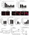
Suppression of IA by S1P1 agonists in rats and non‐human primates. (A, B) Concentration‐dependent suppression of rat IA development by ASP4058. IA was induced in rats administered the indicated doses of ASP4058 p.o. during the whole experimental period as described in the Methods. Specimens of IA induced at the right ACA‐OA bifurcation were prepared at 12 weeks after their induction, and the aneurysm size (A) and the number of macrophages infiltrated into the lesion (B) were examined. Data represent mean ± SEM. Number of animals used is shown in parentheses. (C, D) Suppression of MCP‐1 expression and degenerative changes in the media in IA lesions after treatment with ASP4058. Preparations of IA lesions from vehicle or ASP4058 (0.03 mg·kg−1)‐treated rats were immunostained for MCP‐1 (n = 5 per group) (C, red) and α‐SMA (n = 12 per group) (D, red) with nuclear DAPI staining (blue). Representative images are shown. Boxes in (D) show the region magnified in the respective right panels. Bar, 50 μm. (E, F) Effect of TASP0277308, an S1P1 antagonist, on the aneurysm size and the number of macrophages infiltrated in the lesions. IA was induced in rats administered the indicated doses of TASP0277308, p.o., twice a day during the whole experimental period. Specimens of IA induced at right ACA‐OA bifurcation were prepared at 4 weeks after the induction and the aneurysm size (E) and the number of macrophages in the lesion (F) were then analysed. IA formation was facilitated by 3‐aminopropionitrile treatment in these rats. Data represent mean ± SEM. Number of animals used is shown in parentheses. (G, H) Effects of S1P1 agonists, KRP‐203, BAF312 and fingolimod on the aneurysm size. KRP‐203 and BAF312 were administered at 0.01 and 0.3 mg·kg−1 daily (G) and fingolimod was administered daily at the indicated doses (H) during whole experimental period and the aneurysm size was examined as in (E). IA formation was facilitated by 3‐aminopropionitrile treatment in these rats. Data represent mean ± SEM. Number of animals used is shown in parentheses. * P < 0.05. (I, J) IAs were first induced in rats for 1 week, which was facilitated by 3‐aminopropionitrile treatment, and then treatment with ASP4058 (0.1 mg·kg−1, once a day) was started. At 7 weeks after the induction of the IA, specimens of right ACA‐OA bifurcation were prepared for analysis of aneurysm size. Data represent mean ± SEM. Number of animals used is shown in parentheses. * P < 0.05. BAPN, 3‐aminopropionitrile. (K) Female Macaca fascicularis underwent the aneurysm induction, as described in the Methods, and the effect of ASP4058 (0.1 mg·kg−1, once a day) on IA formation at five bifurcation sites of the intracranial arteries per animal was evaluated at 52 weeks after the induction. Each dot represents the incidence of aneurysm in each animal, and the horizontal bars represent the median of each group (n = 3 in each group).

Similar articles

Cited by

References

    1. Alexander SP, Davenport AP, Kelly E, Marrion N, Peters JA, Benson HE et al. (2015). The concise guide to PHARMACOLOGY 2015/16: G protein‐coupled receptors. Br J Pharmacol 172: 5744–5869. - PMC - PubMed
    1. Aoki T (2015). Inflammation mediates the pathogenesis of cerebral aneurysm and becomes therapeutic target. Neuroimmunol Neuroinflamm 2: 86–92.
    1. Aoki T, Kataoka H, Ishibashi R, Nozaki K, Egashira K, Hashimoto N (2009). Impact of monocyte chemoattractant protein‐1 deficiency on cerebral aneurysm formation. Stroke 40: 942–951. - PubMed
    1. Aoki T, Kataoka H, Shimamura M, Nakagami H, Wakayama K, Moriwaki T et al. (2007). NF‐kappaB is a key mediator of cerebral aneurysm formation. Circulation 116: 2830–2840. - PubMed
    1. Aoki T, Narumiya S (2012). Prostaglandins and chronic inflammation. Trends Pharmacol Sci 33: 304–311. - PubMed

MeSH terms