Review
. 2017 Jun 1;22(10):1805-1829.
doi: 10.2741/4573.

The roles of microglia/macrophages in tumor progression of brain cancer and metastatic disease

Affiliations
Review

The roles of microglia/macrophages in tumor progression of brain cancer and metastatic disease

Shih-Ying Wu et al. Front Biosci (Landmark Ed). .

Abstract

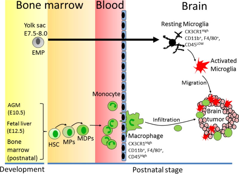
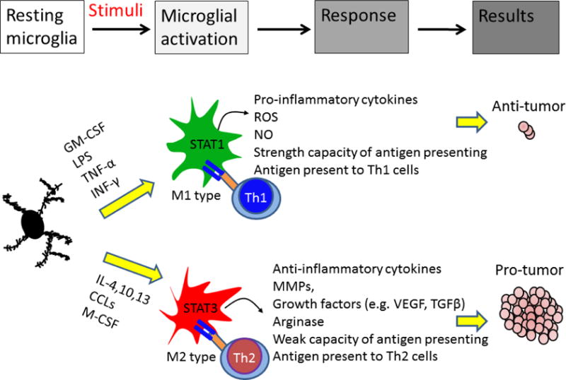
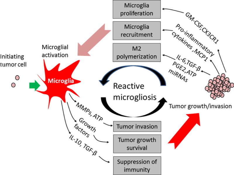
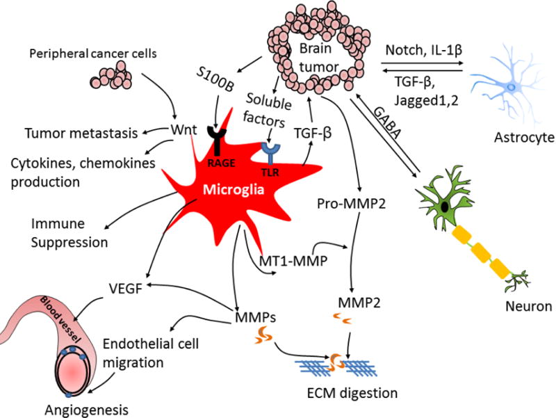
Malignant brain tumors and brain metastases are highly aggressive diseases that are often resistant to treatment. Consequently, the current prognosis of patients with brain tumors and metastases is dismal. Activated microglia and macrophages are often observed in close proximity to or within the malignant tumor masses, suggesting that microglia/macrophages play an important role in brain tumor progression. Microglia, being resident macrophages of the central nervous system, form a major component of the brain immune system. They exhibit anti-tumor functions by phagocytosis and the release of cytotoxic factors. However, these microglia/macrophages can be polarized into becoming tumor-supportive and immunosuppressive cells by certain tumor-derived soluble factors, thereby promoting tumor maintenance and progression. The activated microglia/macrophages also participate in the process of tumor angiogenesis, metastasis, dormancy, and relapse. In this review, we discuss the recent literature on the dual roles of microglia/macrophages in brain tumor progression. We have also reviewed the effect of several well-known microglia/macrophages-derived molecules and signals on brain tumor progression and further discussed the potential therapeutic strategies for targeting the pro-tumor and metastatic functions of microglia/macrophages.


Conflict of interest statement

The authors have declared that no conflict of interest exists.

Figures

Figure 1
Figure 1. Different origin and lineage of microglial cells and macrophages in the brain
Microglial cells derive from erythromyeloid progenitors (EMPs) which locate in the yolk sac (embryonic day 7.5–8.0). In contrast, brain macrophages derive from hematopoietic stem cells (HSCs), which begin at embryonic day 10.5 in the aorta–gonad–mesonephros (AGM) region and at embryonic day 12.5 in the fetal liver. After postnatal stage, HSCs generate monocytes from myeloid precursors (MPs) and macrophage and/or dendritic cell progenitors (MDPs) in the bone marrow. The mature monocytes infiltrate into different oranges and differentiate to macrophage. In brain tumor microenvironment, tumor-released soluble factors recruit microglial cells and macrophages into the tumor site, which promotes tumor growth and metastasis.
Figure 2
Figure 2. Differential roles of activated microglia/macrophages in the brain tumor
Microglia/macrophages have both pro- and anti-tumor potentials. In response to granulocyte-macrophage colony stimulating factor (GM-CSF), lipopolysaccharide (LPS), tumor necrosis factor-α (TNF-α), and interferon-γ (INF-γ) stimuli, microglia/macrophage can be polarized to M1 phenotype. M1 cells exhibit anti-tumor immunity by producing cytotoxic factors and presenting tumor antigen to T helper type 1 cells (Th1) cells. STAT1 activation in M1 cells induces pro-inflammatory cytokines production and increases T-cell-mediated cytolytic activity, leading to tumor cell damage. In response to interleukin-4 (IL-4), chemokine (C-C motif) ligands (CCLs) and macrophage colony-stimulating factor (M-CSF), microglia/macrophage polarize into M2 phenotype. M2 cells express STAT3 that induces anti-inflammatory factors. M2 cells also modulate Th2 cells, which promotes tumor progression. In addition, M2 cells can promote tissue repair and angiogenesis, resulting in tumor progression.
Figure 3
Figure 3. Reactive microgliosis promotes brain tumor progression
Microglial cells become hyper-activated through two mechanisms in brain tumor microenvironment. First, microglial cells become active, produce cytokines, growth factors and matrix-metalloproteases (MMPs) in response to initial tumor cell stimuli. Microglia-secreted factors then promote tumor growth and invasion. Second, tumor cells release growth, chemoattractant, and chemokine factors that recruit and induce another wave of microglial activation, resulting in a perpetuating cycle of microglia activation in the brain tumor. IL-6: Interleukin IL-6, IL-10: Interleukin 10, TGF-β: Transforming growth factor, PGE2: Prostaglandin E2, GM-CSF: granulocyte-macrophage colony stimulating factor, MCP-1: Monocyte chemoattractant protein-1, ATP: Adenosine triphosphate, miRNAs: microRNAs.
Figure 4
Figure 4. Cross-talk between tumor cells and resident cells in the brain
Various secreted soluble factors from tumor cell stimulate microglia and astrocyte activation. The tumor-derived soluble factors bind toll-like receptors (TLRs) on microglia that induces p-38 MAPK activation, resulting in up-regulation of matrix-metalloproteases (MMPs) and membrane type 1-matrix metalloproteinase (MT1-MMP). Microglia-released MMPs then cause extracellular matrix (ECM) digestion that promotes tumor invasion and macrophages/T cells infiltration. In addition, secreted transforming growth factor (TGF-β) from microglial cells triggers the release of pro-MMP2 from tumor cells which is cleaved into active form MMP2 by microglia-released MT1-MMP. Microglia-secreted vascular endothelial growth factor (VEGF) enhances angiogenesis. S100 calcium-binding protein B (S100B) induces receptor for advanced glycation end products (RAGE) activation on microglial cells that induces production of anti-inflammatory cytokines, leading to immune suppression. The metastatic tumor cell induces production of cytokines and chemokines in activated microglia via activation of Wnt signal. Upregulating Wnt signaling in microglia promotes tumor colonization and metastasis. Tumor cells induce astrocyte activation by production of interleukin-1β (IL-1β). Activated astrocytes release TGF-β, Jagged and other factors, which promotes tumor growth and mediates cancer stem cells self-renewal. Neuron-released neurotransmitter gamma-aminobutyric acid (GABA) promotes tumor progression. The interaction of tumor and resident cells induces multiple pathway activation that creates favorable microenvironment for tumor growth. BBB: blood–brain barrier, EGFR: epidermal growth factor receptor, HER: human epidermal growth factor receptor, IL: interleukin, JAG: jagged, MMP: matrix metalloproteinase, TGF: transforming growth factor, VEGF: vascular endothelial growth factor.

References

    1. Gavrilovic IT, Posner JB. Brain metastases: epidemiology and pathophysiology. J Neurooncol. 2005;75:5–14. - PubMed
    1. Tabouret E, Chinot O, Metellus P, Tallet A, Viens P, Goncalves A. Recent trends in epidemiology of brain metastases: an overview. Anticancer Res. 2012;32:4655–62. - PubMed
    1. Polivka J, Jr, Polivka J, Holubec L, Kubikova T, Priban V, Hes O, Pivovarcikova K, Treskova I. Advances in Experimental Targeted Therapy and Immunotherapy for Patients with Glioblastoma Multiforme. Anticancer Res. 2017;37:21–33. - PubMed
    1. Chinot OL, de La Motte Rouge T, Moore N, Zeaiter A, Das A, Phillips H, Modrusan Z, Cloughesy T. AVAglio: Phase 3 trial of bevacizumab plus temozolomide and radiotherapy in newly diagnosed glioblastoma multiforme. Adv Ther. 2011;28:334–40. - PubMed
    1. Staberg M, Michaelsen SR, Rasmussen RD, Villingshoj M, Poulsen HS, Hamerlik P. Inhibition of histone deacetylases sensitizes glioblastoma cells to lomustine. Cell Oncol (Dordr) 2017;40:21–32. - PubMed

Publication types

LinkOut - more resources