. 2017 May 23;8(21):34670-34677.
doi: 10.18632/oncotarget.16668.

Subcloning and characterization of highly metastatic cells derived from human esophageal squamous cell carcinoma KYSE150 cells by in vivo selection

Affiliations

Subcloning and characterization of highly metastatic cells derived from human esophageal squamous cell carcinoma KYSE150 cells by in vivo selection

Masafumi Okuda et al. Oncotarget. .

Abstract

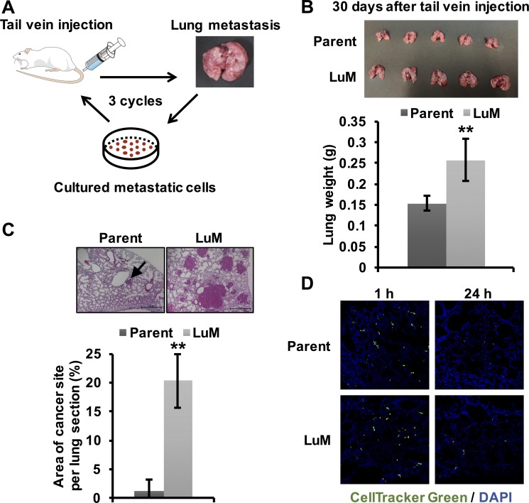
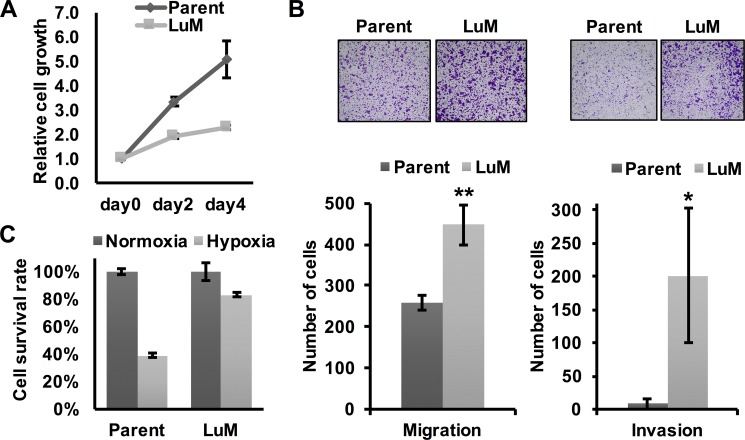
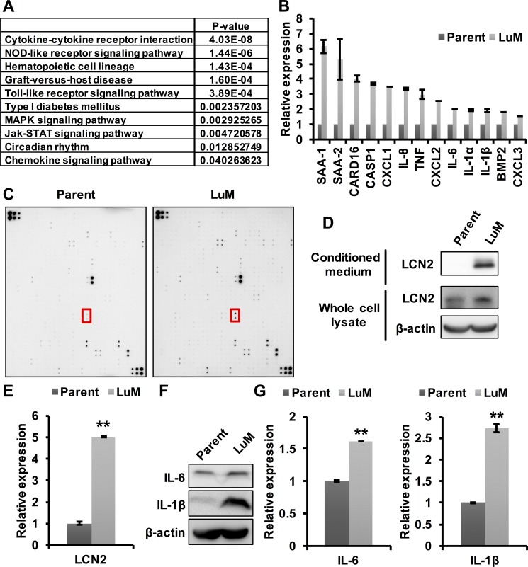
Esophageal cancer is the eighth most common cancer and the sixth most common cause of cancer-related deaths worldwide. Despite the research progress in understanding the disease, the mechanism underlying the metastasis is still unclear. Here, we successfully generated a highly metastatic cell subline, designated as KYSE150-LuM, derived from an esophageal squamous cell carcinoma cell line (KYSE150) by in vivo selection. To elucidate the mechanisms driving metastasis, we characterized the gene expression differences between LuM cells and parent KYSE150 cells. IL-6, IL-1β, and LCN2, previously associated with tumor growth and metastasis, were up-regulated in LuM cells. Recent studies on cancer have increasingly focused on the tumor microenvironment, from which these cytokines are released. The fact that these three cytokines (IL-6, IL-1β, LCN2) were up-regulated in LuM cells indicates that these highly metastatic cells obtained through in vivo selection will be a useful resource for further studies on elucidating the mechanisms underlying the tumor microenvironment which is associated with cytokine-related tumor growth and metastasis. Moreover, LuM cells could disseminate to the lung in shorter period of time in vivo, indicating their utility for in vivo experiments of metastasis and new therapeutic targets in a shorter period of time than currently possible.

Keywords: cytokine; esophageal squamous cell carcinoma (ESCC); in vivo selection; inflammation; lung metastasis.


Conflict of interest statement

CONFLICTS OF INTEREST

There are no conflicts of interest to disclose.

Figures

Figure 1
Figure 1. Generation of highly metastatic cells derived from the KYSE150 cell line
(A) The generation of highly metastatic cells derived from KYSE150, an ESCC cell line, by in vivo selection during the process of lung metastasis. The selection cycle was repeated three times. (B) Ex vivo mouse lung images and weights at 30 days after tail vein injection of parent KYSE150 cells and LuM cells (5.0 × 10⁵ cells in 100 μl of PBS, 5 mice, respectively). Data are represented as mean ± standard deviations. **p < 0.01. (C) Hematoxylin and eosin-stained lung sections and quantification of lung metastatic foci at 30 days after tail vein injection of parent KYSE150 cells and LuM cells (5.0 × 10⁵ cells in 100 μl of PBS, 5 mice, respectively). Scale bar, 500 μm. The arrow indicates metastatic foci. Data are represented as mean ± standard deviations. **p < 0.01. (D) Extravasation assay using immunofluorescent analysis. Cells labeled with CellTracker™ Green were injected into the tail veins of the mice. After a defined time, the mice were sacrificed and frozen sections of the lung were prepared.
Figure 2
Figure 2. Characterization of the highly metastatic cell line LuM
(A) Cell growth curves were determined by crystal violet staining. Cell growth was slower in LuM cells compared with that of the parent KYSE150 cells in vitro. Data are represented as mean ± standard deviations. (B) Cell motility and invasion was assessed by a transwell migration and invasion assay with parent KYSE150 cells or LuM cells. Data are represented as mean ± standard deviations. *p < 0.05. **p < 0.01. (C) Cell survival rates were determined by crystal violet staining. Cell viability under hypoxic conditions (O2 < 0.1%, 48 h) was higher in LuM cells compared with that of parent KYSE150 cells in vitro. Data are represented as mean ± standard deviations.
Figure 3
Figure 3. Involvement of cytokines in the enhanced metastatic ability of LuM cells
(A) Strategy for identifying metastasis-associated genes. In the expression array analysis, 246 genes showed increased expression levels in LuM cells compared with parent KYSE150 cells. Subsequently, gene pathway enrichment analysis showed a strong association of the candidate genes with the cytokine-cytokine receptor pathways. (B) Expression levels of candidate genes by qRT-PCR. Error bars are standard deviations of the means of duplicate experiments. (C) Cytokine array analysis of the cell culture supernatant from parent KYSE150 cells or LuM cells. The red box indicates LCN2. Upper left and lower right on both membrane indicate reference spot. Information of each position on array were shown in manufacturer's instructions. (D) LCN2 protein expression in conditioned medium and the whole cell lysate by western blotting in parent KYSE150 cells and LuM cells. (E) LCN2 secretion levels of the supernatant from parent KYSE150 cells and LuM cells by ELISA. Data are represented as mean ± standard deviations. **p < 0.01. (F) IL-6 and IL-1β protein expression in the whole cell lysate by western blotting in parent KYSE150 cells and LuM cells. (G) IL-6 and IL-1β secretion levels of the supernatant from parent KYSE150 cells and LuM cells by ELISA. Error bars are standard deviations of the means of duplicate experiments. Data are represented as mean ± standard deviations. **p < 0.01.

References

    1. Ferlay J, Soerjomataram I, Dikshit R, Eser S, Mathers C, Rebelo M, Parkin DM, Forman D, Bray F. Cancer incidence and mortality worldwide: sources, methods and major patterns in GLOBOCAN 2012. Int J Cancer. 2015;136:E359–86. doi: 10.1002/ijc.29210. - DOI - PubMed
    1. Lin Y, Totsuka Y, He Y, Kikuchi S, Qiao Y, Ueda J, Wei W, Inoue M, Tanaka H. Epidemiology of esophageal cancer in Japan and China. J Epidemiol. 2013;23:233–42. doi: 10.2188/jea.JE20120162. - DOI - PMC - PubMed
    1. Dotto GP, Rustgi AK. Squamous Cell Cancers: A Unified Perspective on Biology and Genetics. Cancer Cell. 2016;29:622–37. doi: 10.1016/j.ccell.2016.04.004. - DOI - PMC - PubMed
    1. Yokoyama A, Muramatsu T, Omori T, Yokoyama T, Matsushita S, Higuchi S, Maruyama K, Ishii H. Alcohol and aldehyde dehydrogenase gene polymorphisms and oropharyngolaryngeal, esophageal and stomach cancers in Japanese alcoholics. Carcinogenesis. 2001;22:433–9. - PubMed
    1. Muramatsu T, Imoto I, Matsui T, Kozaki K, Haruki S, Sudol M, Shimada Y, Tsuda H, Kawano T, Inazawa J. YAP is a candidate oncogene for esophageal squamous cell carcinoma. Carcinogenesis. 2011;32:389–98. doi: 10.1093/carcin/bgq254. - DOI - PubMed

MeSH terms