. 2017 Apr 14;12(4):e0175369.
doi: 10.1371/journal.pone.0175369. eCollection 2017.

Metabolic changes and inflammation in cultured astrocytes from the 5xFAD mouse model of Alzheimer's disease: Alleviation by pantethine

Affiliations

Metabolic changes and inflammation in cultured astrocytes from the 5xFAD mouse model of Alzheimer's disease: Alleviation by pantethine

Manuel van Gijsel-Bonnello et al. PLoS One. .

Erratum in

Abstract

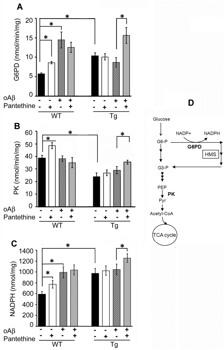
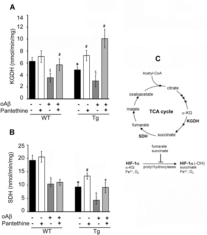
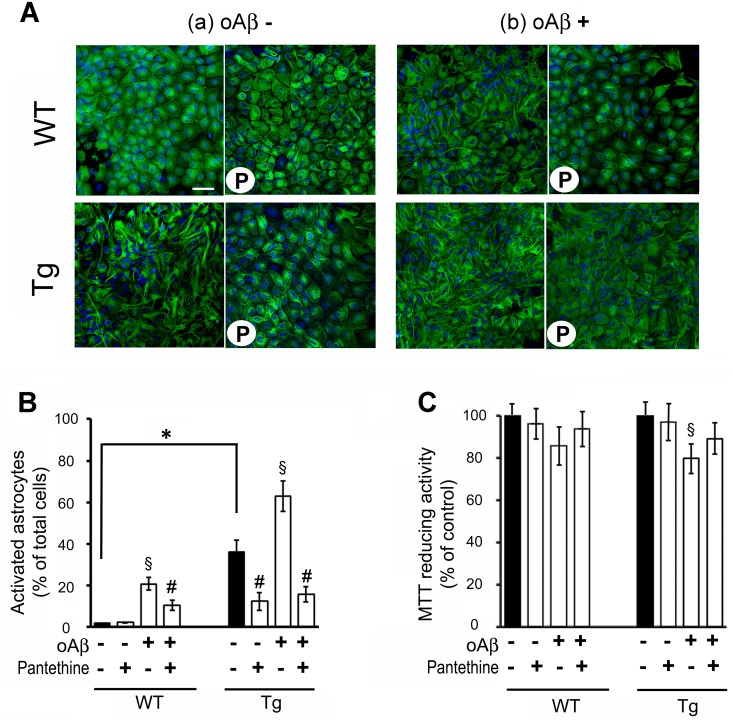
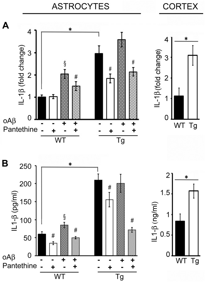
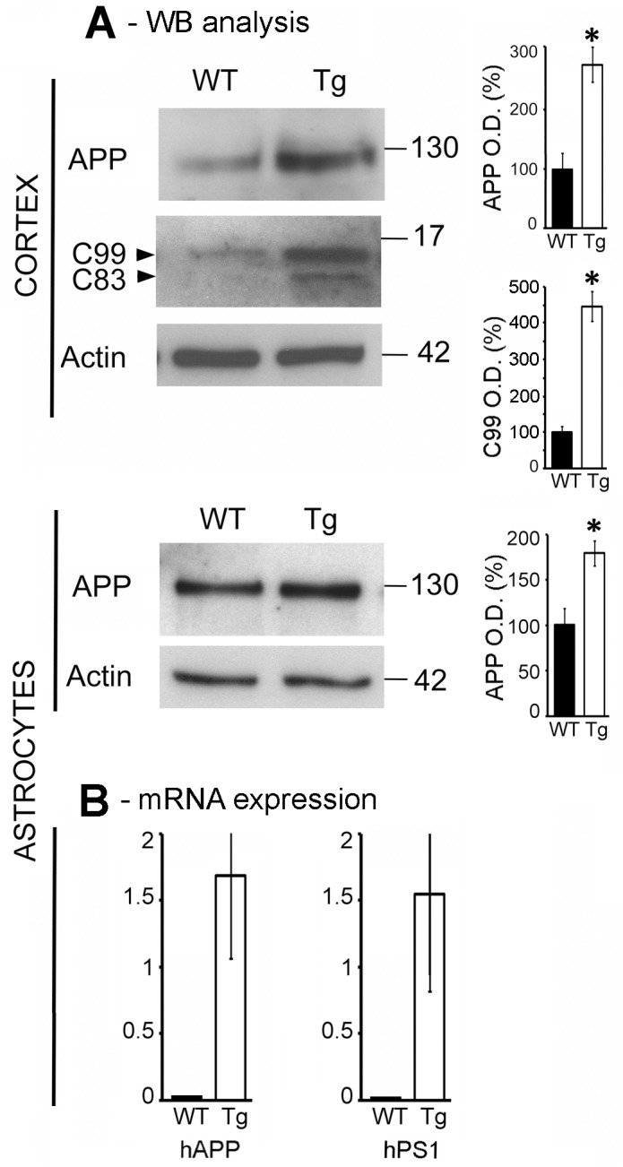
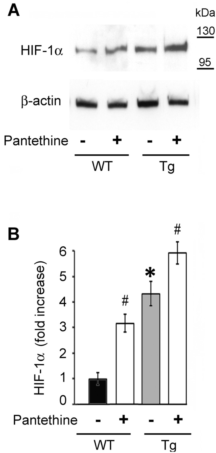
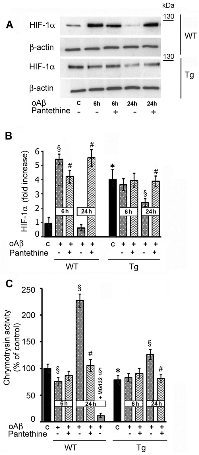
Astrocytes play critical roles in central nervous system homeostasis and support of neuronal function. A better knowledge of their response may both help understand the pathophysiology of Alzheimer's disease (AD) and implement new therapeutic strategies. We used the 5xFAD transgenic mouse model of AD (Tg thereafter) to generate astrocyte cultures and investigate the impact of the genotype on metabolic changes and astrocytes activation. Metabolomic analysis showed that Tg astrocytes exhibited changes in the glycolytic pathway and tricarboxylic acid (TCA) cycle, compared to wild type (WT) cells. Tg astrocytes displayed also a prominent basal inflammatory status, with accentuated reactivity and increased expression of the inflammatory cytokine interleukin-1 beta (IL-1β). Compensatory mechanisms were activated in Tg astrocytes, including: i) the hexose monophosphate shunt with the consequent production of reducing species; ii) the induction of hypoxia inducible factor-1 alpha (HIF-1α), known to protect against amyloid-β (Aβ) toxicity. Such events were associated with the expression by Tg astrocytes of human isoforms of both amyloid precursor protein (APP) and presenilin-1 (PS1). Similar metabolic and inflammatory changes were induced in WT astrocytes by exogenous Aβ peptide. Pantethine, the vitamin B5 precursor, known to be neuroprotective and anti-inflammatory, alleviated the pathological pattern in Tg astrocytes as well as WT astrocytes treated with Aß. In conclusion, our data enlighten the dual pathogenic/protective role of astrocytes in AD pathology and the potential protective role of pantethine.


Conflict of interest statement

Competing Interests: The authors have declared that no competing interests exist.

Figures

Fig 1
Fig 1. Enzymatic activities involved in the glycolytic pathway.
Tg and WT astrocytes were treated or not with pantethine and then exposed or not to exogenous oligomeric Aβ (oAβ). Enzymatic activity and NADPH levels were determined on cell extracts. (A) Glucose-6-phosphate dehydrogenase (G6PD) activity. (B) Pyruvate kinase (PK) activity. (C) NADPH levels. Results are the mean values ± SD from three independent experiments (n = 3 per group). Enzyme activities are expressed as nmol/min/mg protein and NADPH levels are expressed as nmol/mg protein (*, significant difference between experimental groups; p<0.05). (D) Schematic representation of glucose metabolism enlightening the hexose monophosphate shunt (HMS) and PK (G6-P, glucose-6P; G-3P, glyceraldehyde-3P; PEP, phosphoenoylpyruvate; pyr, pyruvate).
Fig 2
Fig 2. Enzymatic activities involved in the TCA cycle.
Tg and WT astrocytes were treated or not with pantethine, then exposed or not to oAβ. Enzymatic activities were determined on mitochondrial preparations. (A) α-Ketoglutarate dehydrogenase (KGDH) activity. (B) succinate dehydrogenase (SDH) activity. Results are the mean values ± SD from three independent experiments (n = 3 per group) and are expressed as nmol/min/mg protein (*, significant difference with the WT group; §, significant difference with the corresponding group not exposed to oAβ; #, significant difference with the corresponding group untreated with pantethine; p<0.05). (C) Schematic representation of the TCA cycle enzymes and metabolites involved in HIF-1α hydroxylation (α-KG: α-ketoglutarate; HIF-1α: hypoxia inducible factor-1α; HIF-1α (-OH): hydroxylated HIF-1α).
Fig 3
Fig 3. Tg and WT astrocyte activation status.
Tg and WT astrocytes were treated or not with pantethine, then exposed or not to oAβ. (A) Confocal microscopy images of astrocyte morphology monitored by GFAP labeling (green); nuclei are labeled with Hoechst (blue). a, Quiescent astrocytes; b, astrocytes exposed to oAβ; P, pantethine treatment. Each picture is representative of images obtained from at least three independent cultures. (B) Astrocyte activation quantified by counting the number of activated cells (stellar shape) as a percent of the total number of cells in 10 randomly selected 105 μm2 fields from all the pictures obtained. (C) Astrocyte viability determined using the MTT assay. The diagrams are mean values ± SD from three independent experiments (n = 3 per group) (*, significant difference between WT and Tg groups; §, significant difference with groups not exposed to oAβ; #, significant difference with the corresponding group untreated with pantethine; p<0.05). Scale bars: 100 μm.
Fig 4
Fig 4. IL-1β expression in WT and Tg astrocytes and cortex.
Tg and WT astrocytes were treated or not with pantethine, then exposed or not to oAβ. IL-1β mRNA expression was determined on cell extracts; cytokine production was assayed on cell supernatants. In cortex samples collected from mice littermates, mRNA expression and protein production were determined on tissue extracts. (A) qPCR analysis. (B) IL-1β ELISA assays. Results are the mean values ± SD from three independent experiments (n = 3 per group); (*, significant difference between Tg and WT groups; §, significant difference with the corresponding group not exposed to oAβ; #, significant difference with the corresponding group untreated with pantethine; p<0.05).
Fig 5
Fig 5. APP is expressed in astrocytes.
(A) Western blot analysis of full-length APP, C99 and C83 CTF fragments in Tg and WT cortices (upper panel) and in astrocytes (bottom panel), using a CTF (C-terminal fragment) antibody specifically recognizing the C-terminal end of APP. Graphs on the right represent the mean ± SD of actin-normalized values of the percentage of variation in optical density (O.D.) relative to WT. (B) mRNA expression levels of human APP (APP) and presenilin-1 (PS1) in Tg and WT astrocytes. Values were obtained from three independent experiments (n = 3 per group) (*, significant difference between Tg and WT groups; p<0.05).
Fig 6
Fig 6. HIF-1α protein levels in Tg and WT astrocytes.
(A) Western blot analysis of HIF-1α expression in astrocytes treated or not with pantethine. (B) Densitometric analysis of western blots. Data were normalized to β-actin and are displayed as fold increase relative to untreated WT astrocytes (black diagram). The results represent the mean ± SD from three independent experiments (n = 3 per group) (*, significant difference with the WT control group; #, significant difference with the corresponding group untreated with pantethine; p<0.05).
Fig 7
Fig 7. Kinetics of HIF-1α protein expression and proteasome activity in Tg and WT astrocytes following oAβ exposure.
(A) Western blot analysis of HIF-1α expression. (B) Densitometry analysis. Data were normalized to β-actin and are displayed as fold increase relative to control untreated WT astrocytes. (C) Proteasomal enzymatic activity assayed using a chymotrypsin substrate. The data are displayed as percent of control untreated WT astrocytes. As a control, enzyme activity was determined in the presence of 0.5 μM of the inhibitor MG132 for 24 h. Bars represent the mean ± SD from three independent experiments (n = 3 per group) (*, significant difference with the corresponding WT control; §, significant difference with the corresponding control; #, significant difference with groups untreated with pantethine; p<0.05).

References

    1. Kalman M (2004) Glia reaction and reactive glia In: Hertz L, editor. Non neuronal cells of the nervous system: function and dysfunction. Amsterdam: Elsevier; pp. 787–835.
    1. Zhao J, O'Connor T, Vassar R. The contribution of activated astrocytes to Abeta production: implications for Alzheimer's disease pathogenesis. J Neuroinflammation. 2011; 8:150 10.1186/1742-2094-8-150 - DOI - PMC - PubMed
    1. Kraft AW, Hu X, Yoon H, Yan P, Xiao Q, Wang Y, et al. Attenuating astrocyte activation accelerates plaque pathogenesis in APP/PS1 mice. FASEB J. 2013; 27:187–198. 10.1096/fj.12-208660 - DOI - PMC - PubMed
    1. Hong HS, Hwang EM, Sim HJ, Cho HJ, Boo JH, Oh SS, et al. Interferon gamma stimulates beta-secretase expression and sAPPbeta production in astrocytes. Biochem Biophys Res Commun. 2003; 307:922–927. - PubMed
    1. Orre M, Kamphuis W, Osborn LM, Jansen AH, Kooijman L, Bossers K, et al. Isolation of glia from Alzheimer's mice reveals inflammation and dysfunction. Neurobiol Aging. 2014; 35:2746–2760. 10.1016/j.neurobiolaging.2014.06.004 - DOI - PubMed

MeSH terms