. 2017 Apr 14;13(4):e1006326.
doi: 10.1371/journal.ppat.1006326. eCollection 2017 Apr.

RIPK3 interacts with MAVS to regulate type I IFN-mediated immunity to Influenza A virus infection

Affiliations

RIPK3 interacts with MAVS to regulate type I IFN-mediated immunity to Influenza A virus infection

Jeffrey Downey et al. PLoS Pathog. .

Abstract

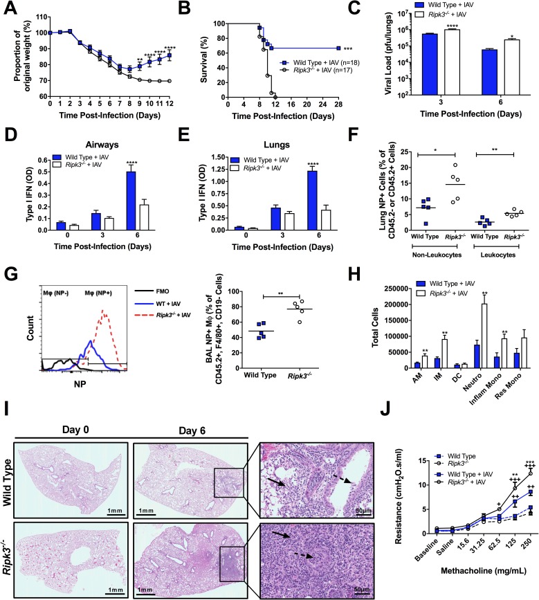
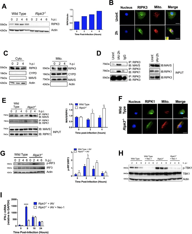
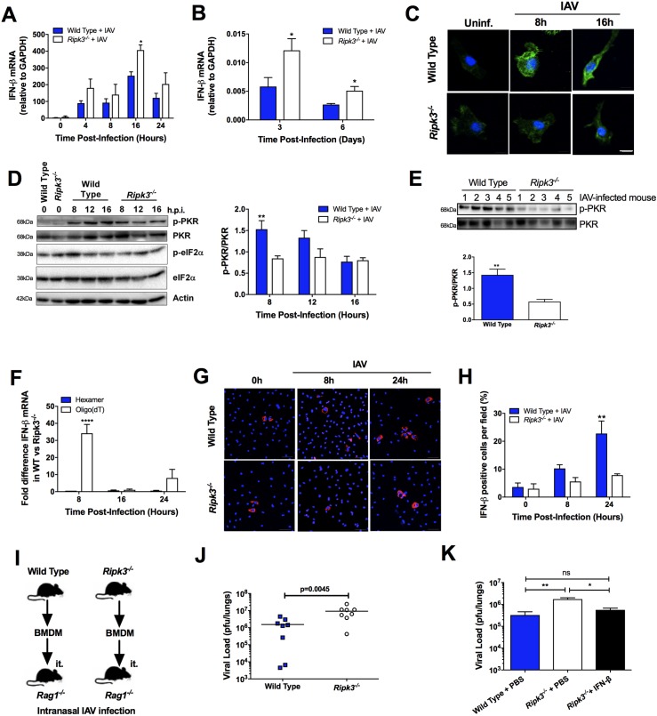
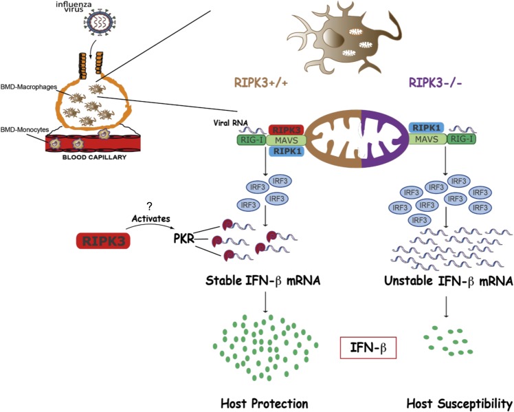
The type I interferon pathway plays a critical role in both host defense and tolerance against viral infection and thus requires refined regulatory mechanisms. RIPK3-mediated necroptosis has been shown to be involved in anti-viral immunity. However, the exact role of RIPK3 in immunity to Influenza A Virus (IAV) is poorly understood. In line with others, we, herein, show that Ripk3-/- mice are highly susceptible to IAV infection, exhibiting elevated pulmonary viral load and heightened morbidity and mortality. Unexpectedly, this susceptibility was linked to an inability of RIKP3-deficient macrophages (Mφ) to produce type I IFN in the lungs of infected mice. In Mφ infected with IAV in vitro, we found that RIPK3 regulates type I IFN both transcriptionally, by interacting with MAVS and limiting RIPK1 interaction with MAVS, and post-transcriptionally, by activating protein kinase R (PKR)-a critical regulator of IFN-β mRNA stability. Collectively, our findings indicate a novel role for RIPK3 in regulating Mφ-mediated type I IFN anti-viral immunity, independent of its conventional role in necroptosis.


Conflict of interest statement

The authors have declared that no competing interests exist.

Figures

Fig 1
Fig 1. RIPK3 restricts early viral replication and prevents excessive inflammation, morbidity, and mortality during IAV infection.
(A-J) WT and Ripk3-/- mice were infected with a sublethal dose (50 pfu) of IAV and morbidity, as a percentage of original weight (A), and survival (B) were assessed. Pulmonary viral loads (C) and total active type I IFN (α and β) via B16-blue reporter cells in the bronchoalveolar lavage (BAL) (D) or lung parenchyma (E) were measured at various times post-infection. (F-G) At 3 days post-infection lungs and BAL from WT and Ripk3-/- mice were collected and cells were intracellularly stained for IAV NP protein. (F) Percentage of NP+ non-leukocytes and leukocytes in the lung. (G) Representative histogram (left panel) of NP protein levels in Mφ (CD45.2+ F4/80+ CD19- cells) of the BAL and the frequency of NP+ Mφ (right panel). (H) Number of alveolar Mφ (AM), interstitial Mφ (IM), dendritic cells (DC), neutrophils (Neutro), Gr1+ inflammatory monocytes (Inflam Mono), and Gr1- resident monocytes (Res Mono) present in the BAL at day 3 post-infection. (I) Micrographs of H&E-stained lung sections prepared prior to and 6 days after IAV infection. At low power, inflammation is absent in both Wild Type and Ripk3-/- (day 0). At high power, the inflammatory infiltrate is composed of lymphocytes, histiocytes and neutrophils within the alveolar space (solid arrow) and bronchiolar lumen (dotted arrow), shown at 6 days post-infection. Scale bar represents 1mm (low magnification) and 50μm (higher magnification). Using flexivent, total respiratory resistance (J) of uninfected or IAV-infected mice was measured following methacholine challenge at day 6 post-infection. Data are represented as mean ± SEM. *p<0.05, **p<0.01, ***p<0.001, ****p<0.0001 between genotypes as indicated, in J, † indicate significant differences over baseline parameter readings of the same genotype. Except in A and B (as indicated), n = 4–8 animals per group per time point.
Fig 2
Fig 2. RIPK3-deficient BMD-Mφ are impaired in anti-viral immunity, independent of the necroptosis pathway.
(A-F, I-J) BMD-Mφ from WT and Ripk3-/- mice were generated and infected with IAV at MOI 1. Total active type I IFN (α and β) (A) and IFN- β (B) was assessed in the supernatants. (C) The relative levels of viral NS1 mRNA were determined via qPCR. (D) BMD-Mφ from WT and Ripk3-/- mice were infected with IAV and the level of viral protein NP was analyzed by flow cytometry. Zebra plots (left panel) are representative of the 24h time-point and numbers adjacent to the gates indicate percent of NP+ Mφ as quantified in the right panel. Level of total active type I IFN in cell culture supernatants (E) and viral load (F) in BMD-Mφ from WT mice treated, or not, with the selective RIPK3 inhibitor GSK‘843 (10μM) and infected with IAV. (G-H) Human monocyte-derived Mφ treated, or not, with the selective RIPK3 inhibitor GSK‘843 (10μM) were infected with IAV H3N2. Levels of active type I IFN (G) and viral load (H) were assessed in culture supernatants 24h after infection. (I) BMD-Mφ from WT and Ripk3-/- mice were generated and treated with various combinations of zVAD-FMK (zVAD, 25μM) and necrostatin-1 (Nec-1, 10μM) for 1h and then were infected with IAV. Necroptosis was assessed by lactate dehydrogenase (LDH) assay after 24h of IAV infection. (J) LDH was measured in BMD-Mφ cell culture supernatants following IAV infection at various time points. Data are representative of the mean ± SEM of triplicate wells and are representative of at least 3 experiments. *p<0.05, **p<0.001, ***p<0.001, ****p<0.0001
Fig 3
Fig 3. RIPK3 interacts with MAVS in IAV-infected BMD-Mφ regulating TBK1/IRF3 dependent type I IFN pathway.
BMD-Mφ were infected with IAV at an MOI of 5 (A-H) or 1 (I). (A) Western blot analysis of RIPK3 expression at various times post-IAV infection in WT BMD-Mφ. Densitometry analysis to quantify ratio of RIPK3 to β-actin is shown in the right panel. (B) Immunofluorescence analysis of co-localization of RIPK3 (green) and mitochondria (red) in WT BMD-Mφ infected or not with IAV. Yellow regions are the areas of RIPK3 and mitochondria colocalization. Nuclei were stained with Hoechst (blue). The scale bars represent 10μm. (C) BMD-Mφ lysates were collected at 0, 2 and 4 hours post-IAV infection. Cytosolic and mitochondrial fractions were isolated and analyzed by western blot for RIPK3 and MAVS. Actin and mitochondrial protein CYPD were used as loading controls and to ensure purity of the fractions. (D-E) BMD-Mφ lysates were collected at 0, 2 (D-E) and 4 (E) hours post-IAV infection and immunoprecipitation was performed with anti-RIPK3 (D) or anti-RIPK1 (E). Samples were then analyzed by immunoblotting for MAVS or RIPK1. (E, right panel) Densitometry analysis to quantify the interaction between MAVS and RIPK1 is shown, representative blot in left panel (n = 3). (F) Immunofluorescence analysis of co-localization of RIPK1 (green) and mitochondria (red) in WT and Ripk3-/- BMD-Mφ 2 hours post IAV-infection. Nuclei were stained with Hoechst (blue). The scale bars represent 10μm. (G) Representative blot (left panel) of phosphorylated IRF3 in WT and Ripk3-/- BMD-Mφ infected, or not, with IAV at various times post-infection. Densitometry analysis to quantify ratio of phosphorylated IRF3 to total IRF3 is shown in the right panel (n = 3). (H-I) WT and Ripk3-/- BMD-Mφ were pretreated with/without necrostatin-1 (Nec-1, 10μM) for 1h and then were infected, or not, with IAV. Representative blot of the phosphorylation of TBK1, determined by western blot as in B. (I) Total RNA was extracted and the expression of IFN-β mRNA was determined by qPCR. Data are expressed as mean ± SEM representative of at least three independent experiments. *p<0.05, **p<0.001, ****p<0.0001.
Fig 4
Fig 4. RIPK3 regulates IFN-β mRNA integrity through activation of PKR.
Total RNA was extracted from IAV-infected BMD-Mφ (MOI 1) (A) or cells of the BAL (50 pfu) (B) and the expression of IFN-β mRNA was determined by qPCR. (C) Phosphorylation of PKR (green) was analyzed by immunofluorescence in WT and Ripk3-/- BMD-Mφ at different time point post IAV-infection. Nuclei were stained with Hoechst (blue). The scale bars represent 10μm. (D) Phosphorylated and total forms of PKR and eIF2α in whole-cell lysates were analysed by immunoblotting. β-Actin was used as a loading control. One representative blot is shown (left panel). Densitometry analysis to quantify the ratio of phosphorylated PKR relative to total PKR (n = 4, right panel). (E) Cells were harvested from the BAL of infected (50 pfu) WT or RIPK3-deficient mice and levels of phosphorylated and total PKR were determined by western blot (top panel). Densitometry analysis to quantify the ratio of phosphorylated PKR relative to total PKR is shown in the bottom panel. (F) Difference in the expression of IFN-β mRNA between WT and Ripk3-/- BMD-Mφ infected with IAV. Gene expression was analyzed by qPCR following cDNA generation using random hexamers (blue bars) or oligo(dT) primers (white bars). (G) Confocal images showing IFN-β production in IAV-infected BMD-Mφ. Cells were stained with a rabbit polyclonal antibody specific for IFN-β (red) as well as nuclear dye Hoechst (blue). (H) Percentage of cells positive for IFN-β per random field. The scale bars represent 50μm. (I) BMD-Mφ (1x10⁶ cells) from WT and Ripk3-/- mice were adoptively transferred (i.t.) into naïve Rag1-/- mice, which were then infected with 500 PFU of IAV 2h post-transfer. (J) Viral load was assessed 3 days after IAV-infection (n = 8, compilation of 2 experiments). (K) Wild Type and Ripk3-/- mice were infected with 50 pfu of IAV. After 2 days, mice were intranasally administered PBS or 2000U of IFN-β. Viral load was determined at day 3 post-infection by standard plaque assay (n = 7–8 mice/group, compilation of 2 experiments). *p<0.05 **p <0.01, ****p <0.0001, ns = not significant.
Fig 5
Fig 5. RIPK3 enhances innate anti-viral immunity against Influenza A virus.
Pulmonary infection by IAV triggers the recruitment of monocytes from the bone marrow that differentiate into macrophages. IAV encounters and infects those macrophages, where viral RNA activates the RIG-I/MAVS pathway, leading to production of the key anti-viral cytokine IFN-β. IAV-induced RIPK3 interaction with MAVS at the mitochondria and may represent an immune evasion strategy to decrease IFN-β production. In the absence of RIPK3, there is increased RIPK1/MAVS interactions, which enhance downstream signaling, resulting in higher TBK1/IRF3 activation and IFN-β mRNA levels. However, this mechanism is counteracted by the RIPK3-mediated activation of PKR. PKR stabilizes IFN-β mRNA through the poly(A) tail, leading to increased IFN-β protein production and, ultimately, host protection.

References

    1. Kumagai Y, Takeuchi O, Kato H, Kumar H, Matsui K, Morii E, et al. Alveolar macrophages are the primary interferon-alpha producer in pulmonary infection with RNA viruses. Immunity. 2007;27(2):240–52. doi: 10.1016/j.immuni.2007.07.013 - DOI - PubMed
    1. Wang J, Nikrad MP, Travanty EA, Zhou B, Phang T, Gao B, et al. Innate immune response of human alveolar macrophages during influenza A infection. PLoS One. 2012;7(3):e29879 Epub 2012 Mar 2. doi: 10.1371/journal.pone.0029879 - DOI - PMC - PubMed
    1. Goritzka M, Makris S, Kausar F, Durant LR, Pereira C, Kumagai Y, et al. Alveolar macrophage-derived type I interferons orchestrate innate immunity to RSV through recruitment of antiviral monocytes. J Exp Med. 2015;212(5):699–714. Epub 2015 Apr 20. doi: 10.1084/jem.20140825 - DOI - PMC - PubMed
    1. Liu S, Cai X, Wu J, Cong Q, Chen X, Li T, et al. Phosphorylation of innate immune adaptor proteins MAVS, STING, and TRIF induces IRF3 activation. Science. 2015;347(6227):aaa2630 doi: 10.1126/science.aaa2630 - DOI - PubMed
    1. Balachandran S, Roberts PC, Brown LE, Truong H, Pattnaik AK, Archer DR, et al. Essential role for the dsRNA-dependent protein kinase PKR in innate immunity to viral infection. Immunity. 2000;13(1):129–41. - PubMed

MeSH terms

Substances