

Mycobacterium ulcerans low infectious dose and mechanical transmission support insect bites and puncturing injuries in the spread of Buruli ulcer
- PMID: 28410412
- PMCID: PMC5406025
- DOI: 10.1371/journal.pntd.0005553
Mycobacterium ulcerans low infectious dose and mechanical transmission support insect bites and puncturing injuries in the spread of Buruli ulcer
Abstract
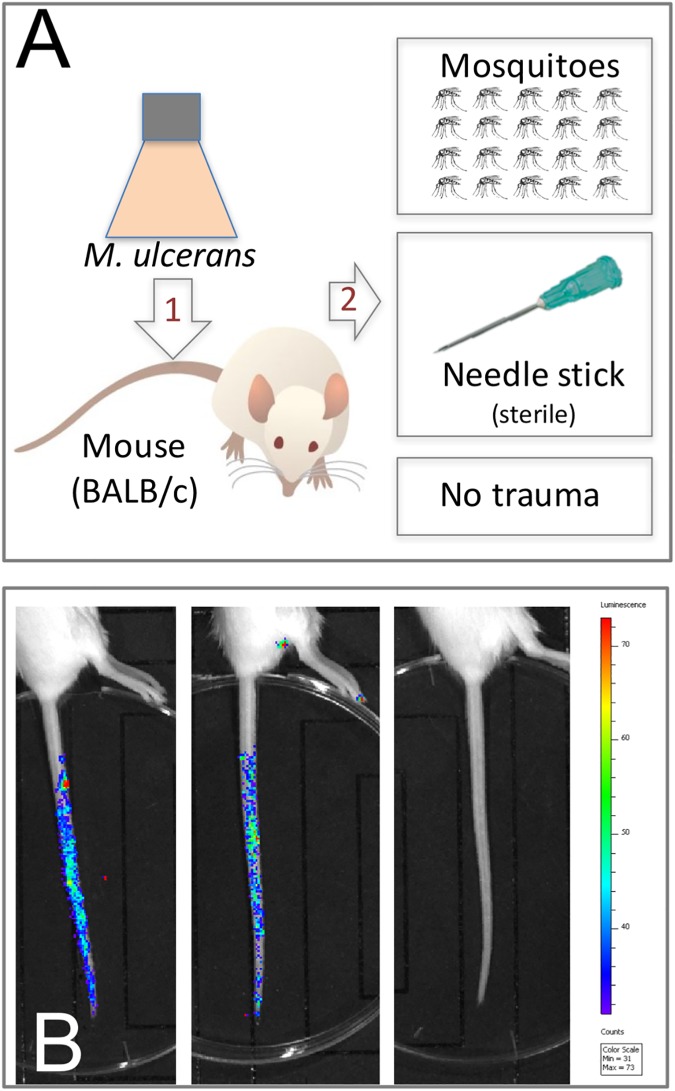
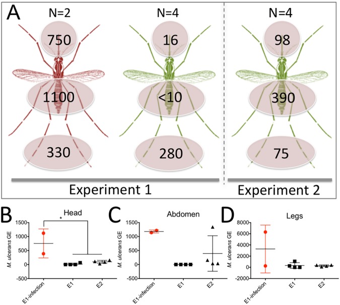
Addressing the transmission enigma of the neglected disease Buruli ulcer (BU) is a World Health Organization priority. In Australia, we have observed an association between mosquitoes harboring the causative agent, Mycobacterium ulcerans, and BU. Here we tested a contaminated skin model of BU transmission by dipping the tails from healthy mice in cultures of the causative agent, Mycobacterium ulcerans. Tails were exposed to mosquito (Aedes notoscriptus and Aedes aegypti) blood feeding or punctured with sterile needles. Two of 12 of mice with M. ulcerans contaminated tails exposed to feeding A. notoscriptus mosquitoes developed BU. There were no mice exposed to A. aegypti that developed BU. Eighty-eight percent of mice (21/24) subjected to contaminated tail needle puncture developed BU. Mouse tails coated only in bacteria did not develop disease. A median incubation time of 12 weeks, consistent with data from human infections, was noted. We then specifically tested the M. ulcerans infectious dose-50 (ID50) in this contaminated skin surface infection model with needle puncture and observed an ID50 of 2.6 colony-forming units. We have uncovered a biologically plausible mechanical transmission mode of BU via natural or anthropogenic skin punctures.
Conflict of interest statement
The authors have declared that no competing interests exist.
Figures

References
-
- Anon. Second WHO report on neglected tropical diseases: sustaining the drive to overcome the global impact of neglected tropical diseases. Geneva, Switzerland: World Health Organization; 2013. p. 11–7.
-
- P. M, Tolhurst JC, Buckle G, Sissons HA. A new mycobacterial infection in man. J Pathol Bacteriol. 1948;60(1):93–122. - PubMed
-
- Johnson PD, Stinear T, Small PL, Pluschke G, Merritt RW, Portaels F, et al. Buruli ulcer (M. ulcerans infection): new insights, new hope for disease control. PLoS Med. 2005;2(4):e108 PubMed Central PMCID: PMC1087202. doi: 10.1371/journal.pmed.0020108 - DOI - PMC - PubMed
-
- Amofah GK, Sagoe-Moses C, Adjei-Acquah C, Frimpong EH. Epidemiology of Buruli ulcer in Amansie West district, Ghana. Trans R Soc Trop Med Hyg. 1993;87(6):644–5. - PubMed
-
- Asiedu K, Etuaful S. Socioeconomic implications of Buruli ulcer in Ghana: a three-year review. Am J Trop Med Hyg. 1998;59(6):1015–22. - PubMed
Publication types
MeSH terms
LinkOut - more resources
Full Text Sources
Other Literature Sources
Medical
Miscellaneous
