Review
. 2017 May;35(2):339-374.
doi: 10.1016/j.ncl.2017.01.008.

Frontotemporal Dementia

Affiliations
Review

Frontotemporal Dementia

Nicholas T Olney et al. Neurol Clin. 2017 May.

Abstract

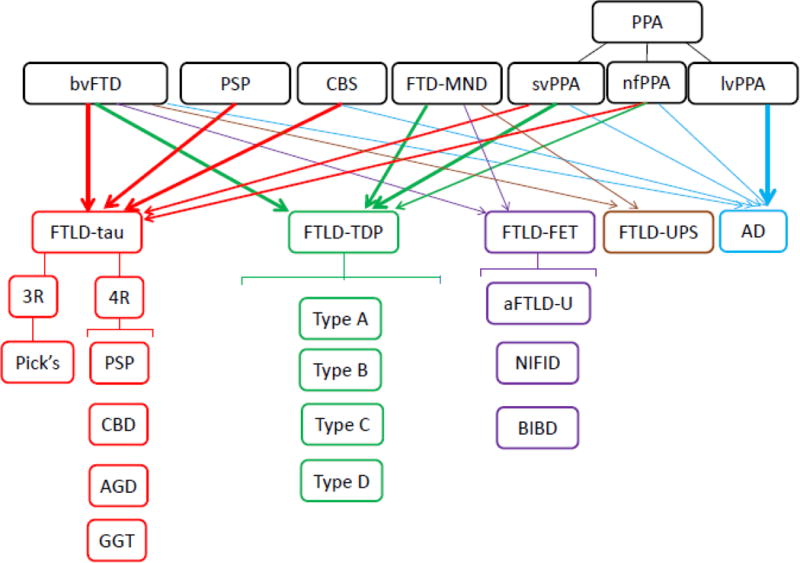
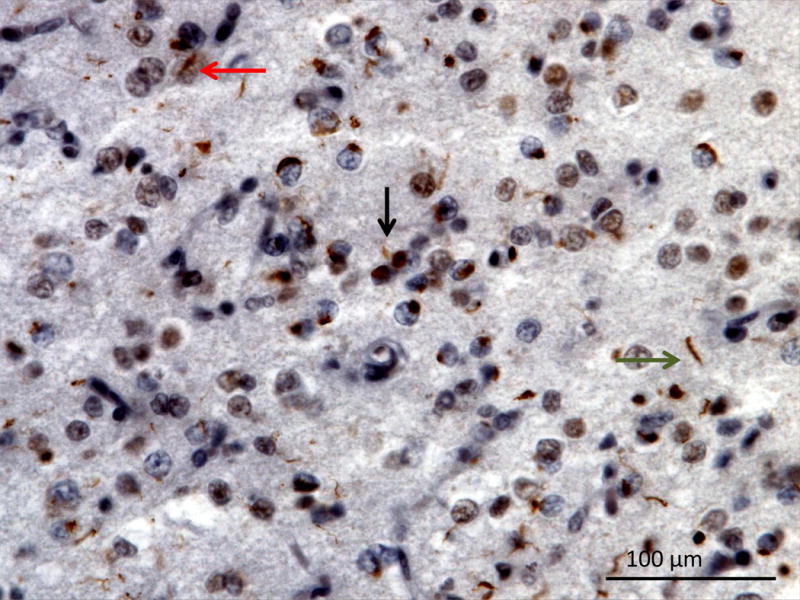
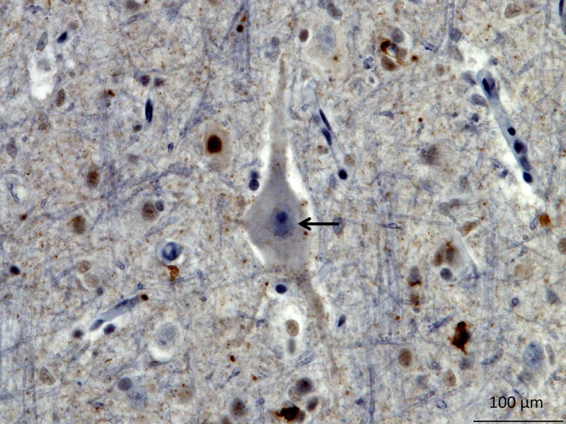
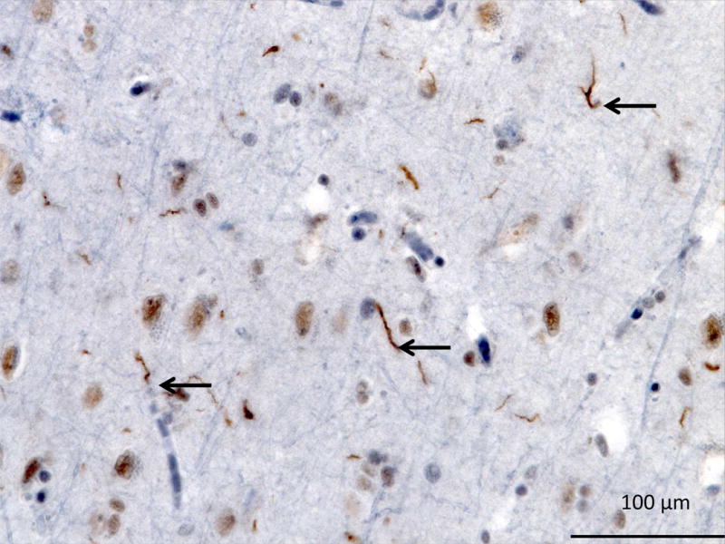
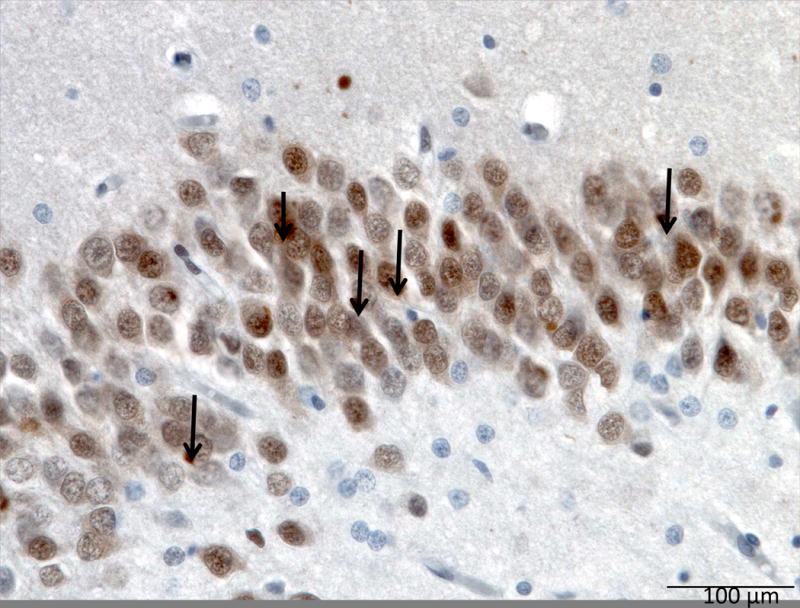
Frontotemporal dementia (FTD) is a heterogeneous disorder with distinct clinical phenotypes associated with multiple neuropathologic entities. Presently, the term FTD encompasses clinical disorders that include changes in behavior, language, executive control, and often motor symptoms. The core FTD spectrum disorders include behavioral variant FTD, nonfluent/agrammatic variant primary progressive aphasia, and semantic variant PPA. Related FTD disorders include frontotemporal dementia with motor neuron disease, progressive supranuclear palsy syndrome, and corticobasal syndrome. In this article, the authors discuss the clinical presentation, diagnostic criteria, neuropathology, genetics, and treatments of these disorders.

Keywords: Corticobasal syndrome (CBS); Frontotemporal dementia (FTD); Motor neuron disease; Nonfluent PPA; Primary progressive aphasia; Progressive supranuclear palsy (PSP); Semantic PPA.


Figures

Figure 1
Figure 1. bvFTD MRI
MRI of a patient with bvFTD showing severe frontal and temporal lobe atrophy. The patient is a 60 year old female who developed symptoms of withdrawal, not taking care of her farm animals, 8 years prior. Her symptoms progressed to impaired hygiene, lack of empathy, and disobeying the rules of the road. In the last 2 years she developed repetitive behaviors, hyperorality, incontinence and a non-fluent aphasia.
Figure 2
Figure 2. Paintings
Paintings made by a 53 year old male with FTLD that had both language and behavioral symptoms. He developed compuslions for painting as one of his early symptoms and it is postulated that as his right temporal lobe become more involved that the faces in his work became less detailed and began to have a more generic smile with teeth.
Figure 3
Figure 3. Left svPPA MRI
MRI showing left temporal lobe atrophy in sagittal, coronal and axial cuts. The patient is a 61 year old man, whose first symptom was 6 years prior with the inability to name apples in a fruit bowl. His problems with semantics and naming progressed and 3 years prior he had difficulty recognizing faces of neighbors. More recent symptoms include the urge to lick people and things (hyperorality).
Figure 4
Figure 4. Right svPPA MRI
MRI shows bitemporal atrophy right greater than left. The patient is a 64 year old woman, whose first symptom was 10 years prior, telling repetitive stories that slowly became less appropriate and embarrassed her friends and family. Six years prior, she developed obsessions of taking specific walking routes to collect cigarette butts. More recently she has shown lack of empathy towards her family and dog.
Figure 5
Figure 5. nfvPPA MRI
MRI showing predominant left posterior fronto-insular atrophy. The patient is an 84 year old woman, whose first symptom was word finding difficulties 7 years prior. Over time she developed impaired grammar and a speech apraxia as her speech output diminished.
Figure 6
Figure 6. Clinical and pathological correlations in FTD spectrum disorders
This figure summarizes the overlap of FTD spectrum disorders (bvFTD, PSP, CBS, FTD-MND, svPPA, nfPPA) and their neuropathology (FTLD-tau, FTLD-TDP, FTLD-FET, FTLD-UPS) with a small portion of clinical syndromes being caused by AD pathology. The clinical syndrome of lvPPA is highly correlated with AD pathology.
Figure 7
Figure 7. 3R tau in Pick’s Disease
Photomicrograph of the middle frontal gyrus from a patient with Pick’s disease. Tau immunohistochemistry demonstrates numerous Pick’s bodies (black arrow) and ballooned neurons or Pick’s cells (red arrows). Image courtesy of Salvatore Spina MD, PhD UCSF
Figure 8
Figure 8. 4R tau in PSP
Tau immunohistochemistry of the superior frontal sulcus showing tufted astrocytes (black arrows), and a globose tangle (red arrow). Image courtesy of Salvatore Spina MD, PhD UCSF
Figure 9
Figure 9. 4R tau in CBD
Tau immunohistochemistry in the paracentral gyrus showing astrocytic plaques (black arrows). Image courtesy of Salvatore Spina MD, PhD UCSF
Figure 10
Figure 10. TDP-43 Type A
Orbitofrontal gyrus showing many short dystrophic neurites (black arrow), neuronal cytoplasmic inclusions (green arrow) and a lentiform neuronal intranuclear inclusions (red arrow). TDP immunohistochemistry. Image courtesy of Lea Grinberg MD, PhD UCSF
Figure 11
Figure 11. TDP-43 Type B
TDP immunohistochemistry of the precentral gyrus showing moderate neuronal cytoplasmic inclusions (black arrow). TDP immunohistochemistry. Image courtesy of Lea Grinberg MD, PhD UCSF
Figure 12
Figure 12. TDP-43 Type C
TDP immunohistochemistry of the insula showing long tortuous dystrophic neurites (black arrows), and few neuronal intranuclear inclusions. Image courtesy of Lea Grinberg MD, PhD UCSF
Figure 13
Figure 13. aFTLD-U binding FUS
FUS immunohistochemistry of the dentate fascia (hippocampus) showing many FUS positive neuronal cytoplasmic inclusions (black arrows). Image courtesy of Lea Grinberg MD, PhD UCSF

References

    1. Ahmed Z, Bigio EH, Budka H, Dickson DW, Ferrer I, Ghetti B, Kovacs GG. Globular glial tauopathies (GGT): consensus recommendations. Acta Neuropathol. 2013;126(4):537–544. doi: 10.1007/s00401-013-1171-0. - DOI - PMC - PubMed
    1. Alzheimer A. Uber eigenartige Krankheitsfalle des spateren Alters. Z Gesamte Neruol Psychiatr. 1911;4:356–385.
    1. Ames D, Cummings JL, Wirshing WC, Quinn B, Mahler M. Repetitive and compulsive behavior in frontal lobe degenerations. Journal of Neuropsychiatry and Clinical Neurosciences. 1994;6(2):100–113. - PubMed
    1. Arai T, Hasegawa M, Akiyama H, Ikeda K, Nonaka T, Mori H, Oda T. TDP-43 is a component of ubiquitin-positive tau-negative inclusions in frontotemporal lobar degeneration and amyotrophic lateral sclerosis. Biochem Biophys Res Commun. 2006;351(3):602–611. - PubMed
    1. Armstrong MJ, Litvan I, Lang AE, Bak TH, Bhatia KP, Borroni B, Weiner WJ. Criteria for the diagnosis of corticobasal degeneration. Neurology. 2013;80(5):496–503. doi: 10.1212/WNL.0b013e31827f0fd1. - DOI - PMC - PubMed

MeSH terms