. 2017 Apr 18;46(4):649-659.
doi: 10.1016/j.immuni.2017.03.016. Epub 2017 Apr 11.

NAIP-NLRC4 Inflammasomes Coordinate Intestinal Epithelial Cell Expulsion with Eicosanoid and IL-18 Release via Activation of Caspase-1 and -8

Affiliations

NAIP-NLRC4 Inflammasomes Coordinate Intestinal Epithelial Cell Expulsion with Eicosanoid and IL-18 Release via Activation of Caspase-1 and -8

Isabella Rauch et al. Immunity. .

Abstract

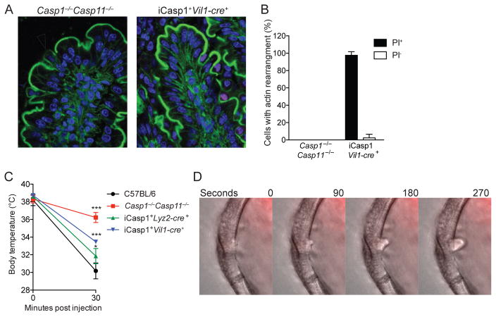
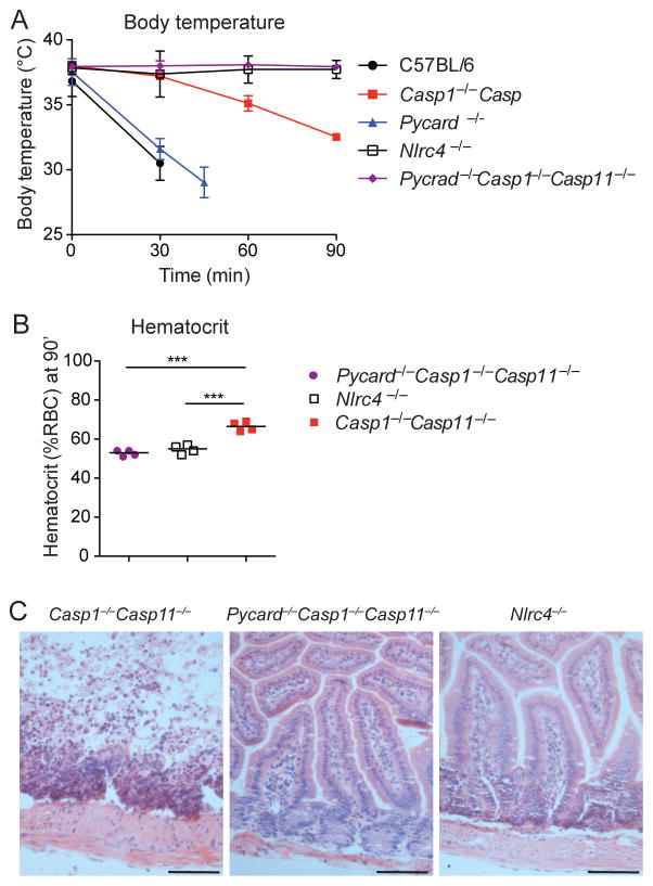
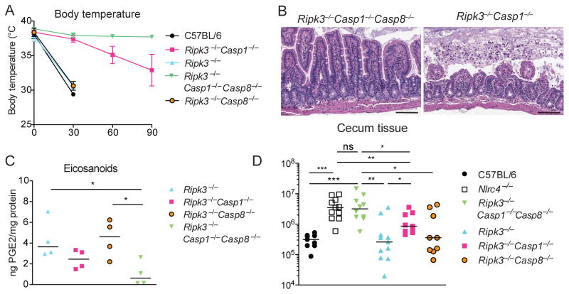
Intestinal epithelial cells (IECs) form a critical barrier against pathogen invasion. By generation of mice in which inflammasome expression is restricted to IECs, we describe a coordinated epithelium-intrinsic inflammasome response in vivo. This response was sufficient to protect against Salmonella tissue invasion and involved a previously reported IEC expulsion that was coordinated with lipid mediator and cytokine production and lytic IEC death. Excessive inflammasome activation in IECs was sufficient to result in diarrhea and pathology. Experiments with IEC organoids demonstrated that IEC expulsion did not require other cell types. IEC expulsion was accompanied by a major actin rearrangement in neighboring cells that maintained epithelium integrity but did not absolutely require Caspase-1 or Gasdermin D. Analysis of Casp1-/-Casp8-/- mice revealed a functional Caspase-8 inflammasome in vivo. Thus, a coordinated IEC-intrinsic, Caspase-1 and -8 inflammasome response plays a key role in intestinal immune defense and pathology.

Keywords: ASC; Caspase-1; Caspase-8; Naip; Nlrc4; expulsion; extrusion; inflammasome; intestinal epithelial cell.


Conflict of interest statement

The authors declare no competing financial interests.

Figures

Figure 1
Figure 1. Specific activation of NLRC4 in intestinal epithelial cells protects from pathogen invasion but can lead to intestinal pathology
(A) Schematic of the Rosa26 locus after successful gene targeting with iNLRC4-IRES-GFP. Yellow triangles represent LoxP sites. (B, C) Mice were injected with 0.8μg/g PA and 0.4μg/g LFn-Fla and monitored for (B) body temperature and (C) hematocrit (30 minutes, n=3). (D, E) Mice were injected with 0.8μg/g PA and 80ng/g LFn-Fla166 and (D) wet/dry ratio of intestinal content and (E) PGE2 amounts of intestinal tissue from mice treated for 30 minutes were determined (n=4–5). (F) Quantification of serum IL-18 using ELISA in mice treated as in B), 60 minute timepoint (n=3). (G) H&E staining of small intestinal tissue from mice treated as in B), 60 minutes post injection. Scale bar = 100μm (H) CFU in cecum 18h after oral S.Typhimurium infection (n=5). Data are representative of 3 independent experiments. (B,F) mean± SD, (C–E,H) median, (B–E) unpaired t-test (in B compared to Nlrc4−/−), (H) Mann-Whitney test, *p<0.01, **p<0.005, ***p<0.001. Please see also Figure S1
Figure 2
Figure 2. Cell autonomous expulsion and lytic death of IECs
(A) Wild-type small intestinal organoid treated with 16μg/ml of PA and 1μg/ml LFn-Fla166 (upper lane) or 100ng/ml TNF-α (lower lane) and stained with propidium iodide. (B) Blinded quantification of C57BL/6 small intestinal organoid cells positive for propidium iodide before expulsion in Fla166Tox vs. TNF-α treatment as in A). n=5–6 movies/ treatment. (C) Propidium iodide and fluorescent phalloidin (actin) or (D) Propidium iodide and GFP in small intestines from mice treated with 0.2μg/g of PA and 0.1μg/g LFn-Fla for 60 minutes. Arrows indicate expulsing cells. Scale bar = 40 (C) 20 (D) μm (E) Quantification of GFP signal in propidium iodide positive and neighboring negative cells from samples as in (D) (n=3). (F) Quantification of actin purse strings in propidium iodide positive cells from samples as in (C) (n=7) (G) Wild-type small intestinal organoid treated with 16μg/ml of PA, 1μg/ml LFn-Fla166 and 2.5μM cytochalasin D and stained with propidium iodide. Data representative of at least 2 independent experiments, unpaired t-test, **p<0.005, ***p<0.001. Please see also Figure S2
Figure 3
Figure 3. Caspase-1 is required for IEC pyroptosis but not IEC expulsion
(A) Propidium iodide and fluorescent phalloidin (actin) in small intestines from mice treated with 0.8μg/g PA and 0.4μg/g LFn-Fla for 60 minutes. Arrows indicate expulsing cells. Scale bar = 20μm (B) Blinded quantification of propidium iodide positive and negative cells with actin purse strings in samples as in (A) (n=3) (C) Mice were treated as in (A) and monitored for body temperature (n=3). (D) Casp1−/− small intestinal organoid treated with 16μg/ml of PA and 1μg/ml LFn-Fla166 and stained with propidium iodide. Data representative of at least 2 independent experiments. (B, C): mean± SD, (C) unpaired t-test compared to Casp1−/−Casp11−/−, ***p<0.001. Please see also Figure S3
Figure 4
Figure 4. Gasdermin D is required for IEC pyroptosis but not IEC expulsion
(A) Propidium iodide and fluorescent phalloidin (actin) in small intestines from mice treated with 0.8μg/g PA and 0.4μg/g LFn-Fla for 60 minutes. Arrows indicate expulsing cells. Scale bar = 20μm (B) Blinded quantification of propidium iodide positive and negative cells with actin purse strings in samples as in (A) (n=3) (C) Mice were injected with 0.8μg/g PA and 0.4μg/g LFn-Fla and monitored for body temperature (n=3). (D) H&E staining of small intestinal tissue from mice treated as in A, 90 minute timepoint. Scale bar = 100μm Data representative of at least 2 independent experiments. (B, C): mean± SD, (C) unpaired t-test compared to Nlrc4−/−,**p<0.005, ***p<0.001. Please see also Figure S4
Figure 5
Figure 5. ASC is required for the Caspase1-independent NLRC4 signaling
(A, B) Mice were injected with 0.8μg/g PA and 0.4μg/g LFn-Fla and monitored for (A) body temperature and (B) hematocrit (90 minutes, n=3). (C) H&E staining of small intestinal tissue from mice treated as in (A), 90 minute timepoint. Scale bar = 100μm Data representative of 2 independent experiments. (A): mean± SD, (B): median. unpaired t-test (in A compared to Nlrc4−/−),**p<0.005, ***p<0.001. Please see also Figure S3
Figure 6
Figure 6. A Caspase 8 inflammasome compensates for loss of Caspase 1
(A) Mice were injected with 0.8μg/g PA and 0.4μg/g LFn-Fla and monitored for body temperature (n=3), mean± SD. (B) H&E staining of small intestinal tissue from mice treated as in (A), 90 minute timepoint. Scale bar = 100μm (C) PGE2 lamounts of intestinal tissue from mice treated as in (A) for 30 minutes were determined (n=4/group), median. Data representative of 2 independent experiments. (D) CFU in cecum 18h after oral S.Typhimurium infection (n=10), combined data of 2 independent experiments. (A+C) unpaired t-test (in A compared to Ripk3−/−Casp1−/−Casp8−/−). (D) Mann-Whitney test, *p<0.01, **p<0.005, ***p<0.001. Please see also Figure S5+6

Comment in

References

    1. Aglietti RA, Estevez A, Gupta A, Ramirez MG, Liu PS, Kayagaki N, Ciferri C, Dixit VM, Dueber EC. GsdmD p30 elicited by caspase-11 during pyroptosis forms pores in membranes. Proc Natl Acad Sci USA. 2016;113(28):7858–63. - PMC - PubMed
    1. Allam R, Maillard MH, Tardivel A, Chennupati V, Bega H, Yu CW, Velin D, Schneider P, Maslowski KM. Epithelial NAIPs protect against colonic tumorigenesis. J Exp Med. 2015;212:369–383. - PMC - PubMed
    1. Anand PK, Malireddi RKS, Lukens JR, Vogel P, Bertin J, Lamkanfi M, Kanneganti TD. NLRP6 negatively regulates innate immunity and host defence against bacterial pathogens. Nature. 2012;488:389–393. - PMC - PubMed
    1. Antonopoulos C, Russo HM, El-Sanadi C, Martin BN, Li X, Kaiser WJ, Mocarski ES, Dubyak GR. Caspase-8 as an Effector and Regulator of NLRP3 Inflammasome Signaling. J Biol Chem. 2015;290:20167–20184. - PMC - PubMed
    1. Ballard JD, Collier RJ, Starnbach MN. Anthrax toxin-mediated delivery of a cytotoxic T-cell epitope in vivo. Proc Natl Acad Sci USA. 1996;93:12531–12534. - PMC - PubMed

MeSH terms