. 2017 Jul 15:155:503-512.
doi: 10.1016/j.neuroimage.2017.04.016. Epub 2017 Apr 12.

A graded tractographic parcellation of the temporal lobe

Affiliations

A graded tractographic parcellation of the temporal lobe

Claude J Bajada et al. Neuroimage. .

Abstract

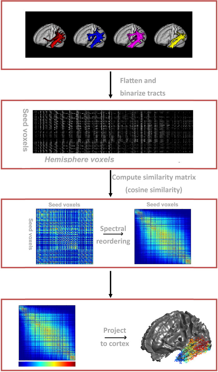
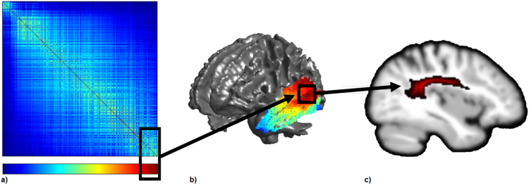
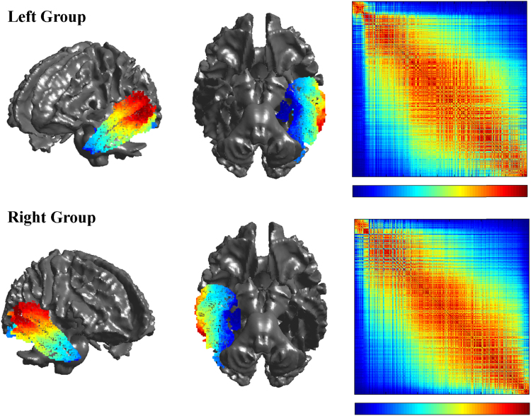
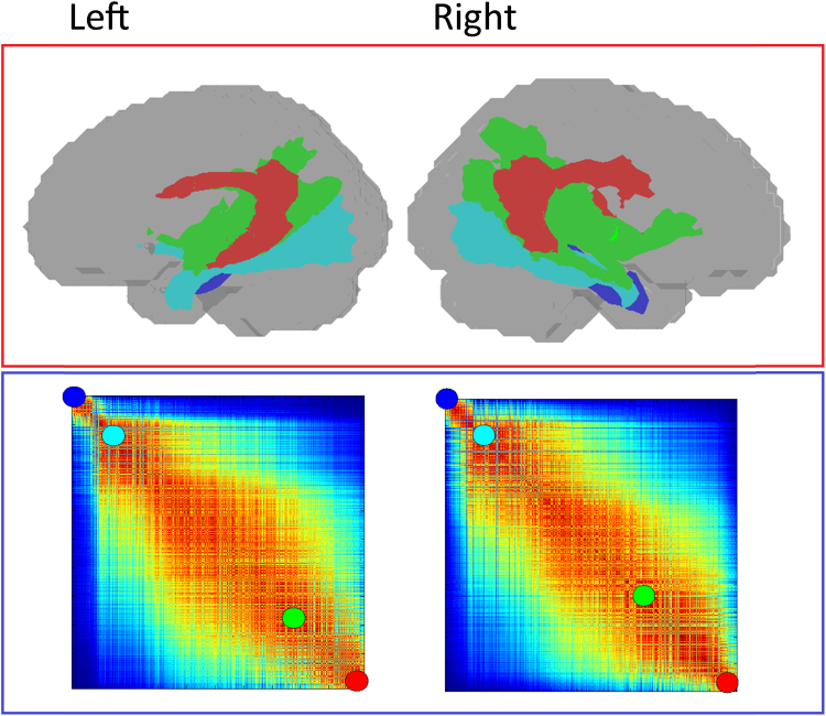
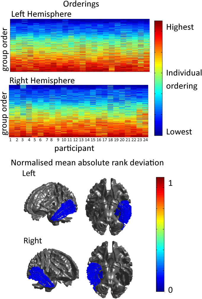
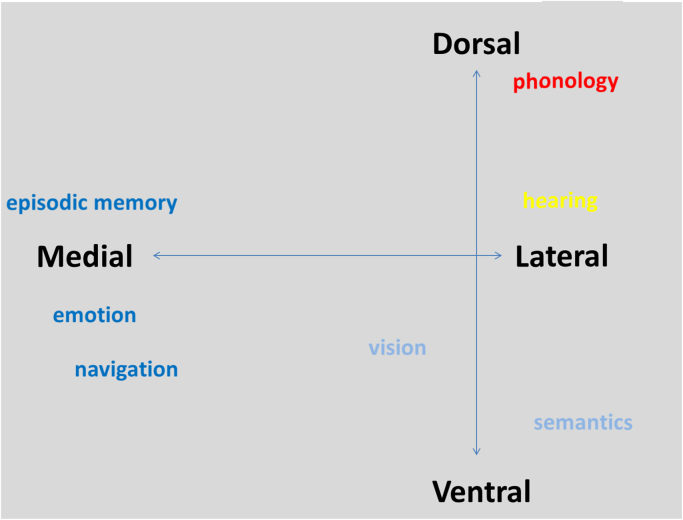
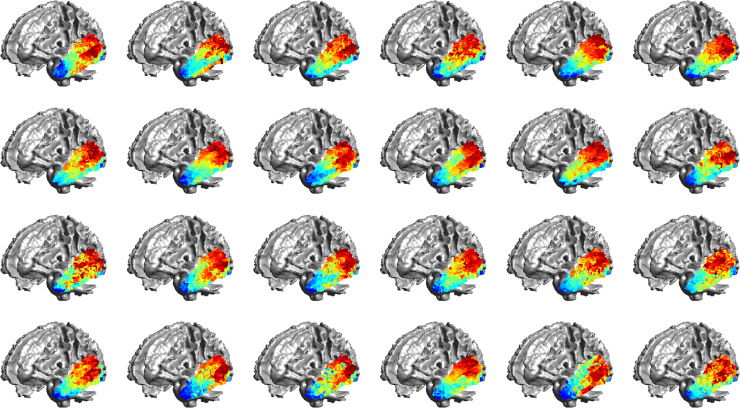
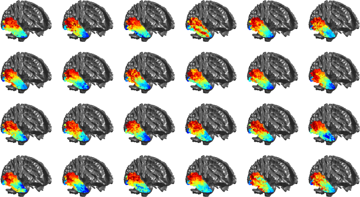
The temporal lobe has been implicated in multiple cognitive domains through lesion studies as well as cognitive neuroimaging research. There has been a recent increased interest in the structural and connective architecture that underlies these functions. However there has not yet been a comprehensive exploration of the patterns of connectivity that appear across the temporal lobe. This article uses a data driven, spectral reordering approach in order to understand the general axes of structural connectivity within the temporal lobe. Two important findings emerge from the study. Firstly, the temporal lobe's overarching patterns of connectivity are organised along two key structural axes: medial to lateral and anteroventral to posterodorsal, mirroring findings in the functional literature. Secondly, the connective organisation of the temporal lobe is graded and transitional; this is reminiscent of the original work of 19th Century neuroanatomists, who posited the existence of some regions which transitioned between one another in a graded fashion. While regions with unique connectivity exist, the boundaries between these are not always sharp. Instead there are zones of graded connectivity reflecting the influence and overlap of shared connectivity.

Keywords: Connectivity based parcellation; Diffusion MRI; Probabilistic tractography; Spectral reordering; Temporal lobe.


Figures

Fig. 1
Fig. 1
–The pipeline to create a graded parcellation of an individual temporal lobe. a) Sample of connectivity profiles from adjacent temporal seed voxels. b) Connectivity profiles are flattened and concatenated to one another to create a 2D matrix representing connectivity of seeds voxels to other voxels. c) A pairwise similarity algorithm is run on b) to generate a similarity matrix. The similarity matrix is spectrally reordered such that seed voxels with strong similarity between their patterns of connectivity are positioned together. d) The reordered matrix is projected onto the brain to visualise the resulting graded parcellation.
Fig. 2
Fig. 2
An illustration of how the underlying connectivity of the graded parcellation can be explored. The black boxes indicate corresponding region in the matrix (a) and their approximate location on the cortex (b). The colour bar under the matrix represents the mapping of colour from the matrix to the cortex. This example shows the far right hand corner of the matrix represented at the red end of the colour bar, which maps onto the posterior superior and middle temporal gyri. A sagittal slice through the underlying tract(s) from a voxel in this region is displayed in panel c.
Fig. 3
Fig. 3
– Group level graded parcellation results of the left and right temporal lobes, displaying lateral and ventral views projected onto the brain as well as the spectrally reordered matrices on which they are based. The colour bar below the matrices has two representations: 1) the level of similarity between left (blue) – right (red), 2) a mapping between the seed in matrix space and in brain space (for example the left most seed (column) in the matrix is coloured blue on the brain while the right most seed is coloured red).
Fig. 4
Fig. 4
Sample connectivity profiles underlying the group reordering for the left and right hemispheres. Four group connectivity profiles were selected at the extremes of the matrix and within the graded section to depict the general order of connectivity profiles. The left and right hemisphere show a similar trend of fibres where early in the ordering the cingulum is prominent followed by more intra-lobar and temporo-frontal tracts. The other end of the matrix is dominated by the temporo-parietal middle longitudinal fasciculus and the arcuate fasciculus.
Fig. 5
Fig. 5
Upper Panel: The individual orderings across all the individual 24 participants. The colours represent the individual's ordering while the y-axis represents the group ordering. Lower Panel: the normalised mean absolute rank deviation for each voxel plotted on the cortex. All voxels have very low mean absolute rank deviations with the lowest being in the medial temporal lobe and the dorsolateral temporal lobe.
Fig. 6
Fig. 6
A schematic showing the structural axes of connectivity and putative cognitive functions that map onto these axes.
Fig. S1
Fig. S1
Individual level graded parcellation results of the left temporal lobe projected onto the brain for each of the 24 participants. See Fig. 3 of main article for group level results and explanation of colour coding.
Fig. S2
Fig. S2
Individual level graded parcellation results of the right temporal lobe projected onto the brain for each of the 24 participants. See Fig. 3 of main article for group level results and explanation of colour coding.

Similar articles

Cited by

References

    1. Amunts K., Zilles K. Architectonic mapping of the human brain beyond Brodmann. Neuron. 2015;88:1086–1107. - PubMed
    1. Anwander A., Tittgemeyer M., Cramon D.Yv, Friederici A.D., Knösche T.R. Connectivity-based parcellation of Broca's area. Cereb. Cortex. 2006;17:816–825. - PubMed
    1. Ashburner J. A fast diffeomorphic image registration algorithm. NeuroImage. 2007;38:95–113. - PubMed
    1. Axer H., Klingner C.M., Prescher A. Fiber anatomy of dorsal and ventral language streams. Brain Lang. 2013;127:192–204. - PubMed
    1. Azadbakht H., Parkes L.M., Haroon H.A., Augath M., Logothetis N.K., de Crespigny A., D'Arceuil H.E., Parker G.J. Validation of high-resolution tractography against in vivo tracing in the macaque visual cortex. Cereb. Cortex. 2015;45:131–136. - PMC - PubMed

Publication types