

Intrinsic membrane properties and cholinergic modulation of mouse basal forebrain glutamatergic neurons in vitro
- PMID: 28411158
- PMCID: PMC5505269
- DOI: 10.1016/j.neuroscience.2017.04.002
Intrinsic membrane properties and cholinergic modulation of mouse basal forebrain glutamatergic neurons in vitro
Abstract
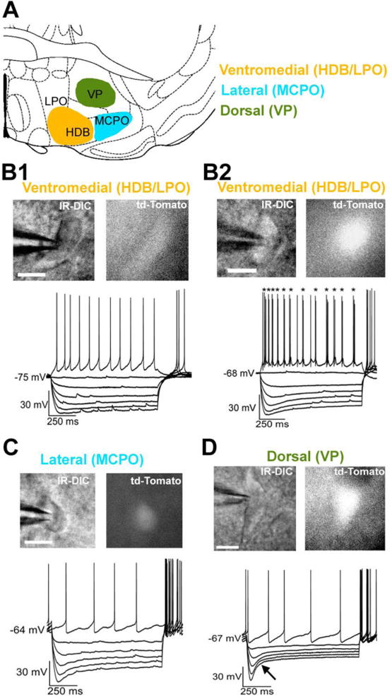
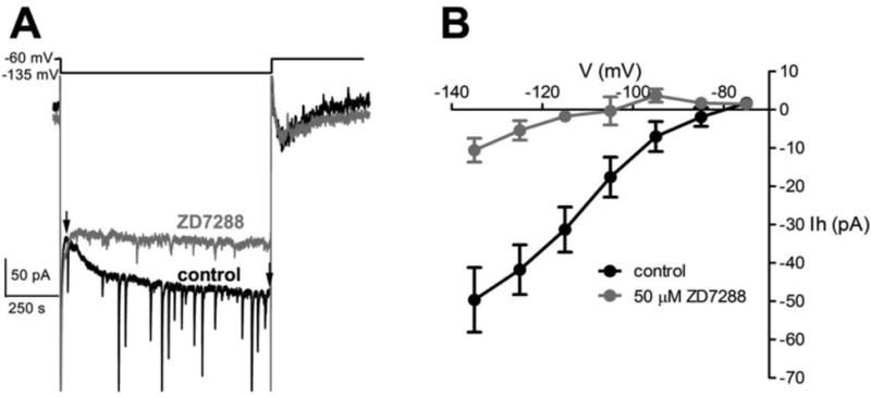
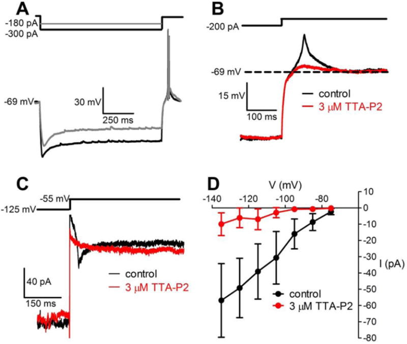
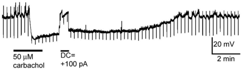
The basal forebrain (BF) controls sleep-wake cycles, attention and reward processing. Compared to cholinergic and GABAergic neurons, BF glutamatergic neurons are less well understood, due to difficulties in identification. Here, we use vesicular glutamate transporter 2 (vGluT2)-tdTomato mice, expressing a red fluorescent protein (tdTomato) in the major group of BF glutamatergic neurons (vGluT2+) to characterize their intrinsic electrical properties and cholinergic modulation. Whole-cell, patch-clamp recordings were made from vGluT2+ neurons in coronal BF slices. Most BF vGluT2+ neurons were small/medium sized (<20µm), exhibited moderately sized H-currents and had a maximal firing frequency of ∼50Hz. However, vGluT2+ neurons in dorsal BF (ventral pallidum) had larger H-currents and a higher maximal firing rate (83Hz). A subset of BF vGluT2+ neurons exhibited burst/cluster firing. Most vGluT2+ neurons had low-threshold calcium spikes/currents. vGluT2+ neurons located in ventromedial regions of BF (in or adjacent to the horizontal limb of the diagonal band) were strongly hyperpolarized by the cholinergic agonist, carbachol, a finding apparently in conflict with their increased discharge during wakefulness/REM sleep and hypothesized role in wake-promotion. In contrast, most vGluT2+ neurons located in lateral BF (magnocellular preoptic area) or dorsal BF did not respond to carbachol. Our results suggest that BF glutamatergic neurons are heterogeneous and have morphological, electrical and pharmacological properties which distinguish them from BF cholinergic and GABAergic neurons. A subset of vGluT2+ neurons, possibly those neurons which project to reward-related areas such as the habenula, are hyperpolarized by cholinergic inputs, which may cause phasic inhibition during reward-related events.
Keywords: Alzheimer’s disease; cortical activation; patch-clamp; sleep; vesicular glutamate transporter; whole-cell.
Copyright © 2017 IBRO. All rights reserved.
Figures
Similar articles
-
Characterization of basal forebrain glutamate neurons suggests a role in control of arousal and avoidance behavior.Brain Struct Funct. 2021 Jul;226(6):1755-1778. doi: 10.1007/s00429-021-02288-7. Epub 2021 May 16. Brain Struct Funct. 2021. PMID: 33997911 Free PMC article.
-
Cholinergic neurons excite cortically projecting basal forebrain GABAergic neurons.J Neurosci. 2014 Feb 19;34(8):2832-44. doi: 10.1523/JNEUROSCI.3235-13.2014. J Neurosci. 2014. PMID: 24553925 Free PMC article.
-
Glutamatergic Ventral Pallidal Neurons Modulate Activity of the Habenula-Tegmental Circuitry and Constrain Reward Seeking.Biol Psychiatry. 2018 Jun 15;83(12):1012-1023. doi: 10.1016/j.biopsych.2018.01.003. Epub 2018 Jan 12. Biol Psychiatry. 2018. PMID: 29452828 Free PMC article.
-
[Advances in the study of the effect of basal forebrain on sleep-wake regulation].Yao Xue Xue Bao. 2016 Aug;51(8):1196-201. Yao Xue Xue Bao. 2016. PMID: 29897712 Review. Chinese.
-
Magnocellular nuclei of the basal forebrain: substrates of sleep and arousal regulation.Sleep. 1995 Jul;18(6):478-500. doi: 10.1093/sleep/18.6.478. Sleep. 1995. PMID: 7481420 Review.
Cited by
-
Basal forebrain contributes to default mode network regulation.Proc Natl Acad Sci U S A. 2018 Feb 6;115(6):1352-1357. doi: 10.1073/pnas.1712431115. Epub 2018 Jan 23. Proc Natl Acad Sci U S A. 2018. PMID: 29363595 Free PMC article.
-
[Changes of membrane properties and synaptic stability of rat retinal ganglion cells during postnatal development].Nan Fang Yi Ke Da Xue Xue Bao. 2018 Aug 30;38(9):1100-1106. doi: 10.12122/j.issn.1673-4254.2018.09.13. Nan Fang Yi Ke Da Xue Xue Bao. 2018. PMID: 30377110 Free PMC article. Chinese.
-
Cholinergic modulation supports dynamic switching of resting state networks through selective DMN suppression.PLoS Comput Biol. 2024 Jun 6;20(6):e1012099. doi: 10.1371/journal.pcbi.1012099. eCollection 2024 Jun. PLoS Comput Biol. 2024. PMID: 38843298 Free PMC article.
-
Characterization of basal forebrain glutamate neurons suggests a role in control of arousal and avoidance behavior.Brain Struct Funct. 2021 Jul;226(6):1755-1778. doi: 10.1007/s00429-021-02288-7. Epub 2021 May 16. Brain Struct Funct. 2021. PMID: 33997911 Free PMC article.
-
Opponent control of behavioral reinforcement by inhibitory and excitatory projections from the ventral pallidum.Nat Commun. 2018 Feb 27;9(1):849. doi: 10.1038/s41467-018-03125-y. Nat Commun. 2018. PMID: 29487284 Free PMC article.
References
-
- Alonso A, Khateb A, Fort P, Jones BE, Muhlethaler M. Differential oscillatory properties of cholinergic and noncholinergic nucleus basalis neurons in guinea pig brain slice. Eur J Neurosci. 1996;8:169–182. - PubMed
Publication types
MeSH terms
Substances
Grants and funding
LinkOut - more resources
Full Text Sources
Other Literature Sources
Miscellaneous
