. 2017 Jun 2;292(22):9117-9135.
doi: 10.1074/jbc.M117.778977. Epub 2017 Apr 14.

2-Alkylquinolone alkaloid biosynthesis in the medicinal plant Evodia rutaecarpa involves collaboration of two novel type III polyketide synthases

Affiliations

2-Alkylquinolone alkaloid biosynthesis in the medicinal plant Evodia rutaecarpa involves collaboration of two novel type III polyketide synthases

Takashi Matsui et al. J Biol Chem. .

Abstract

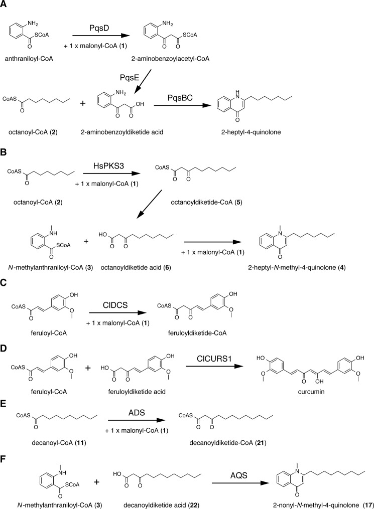
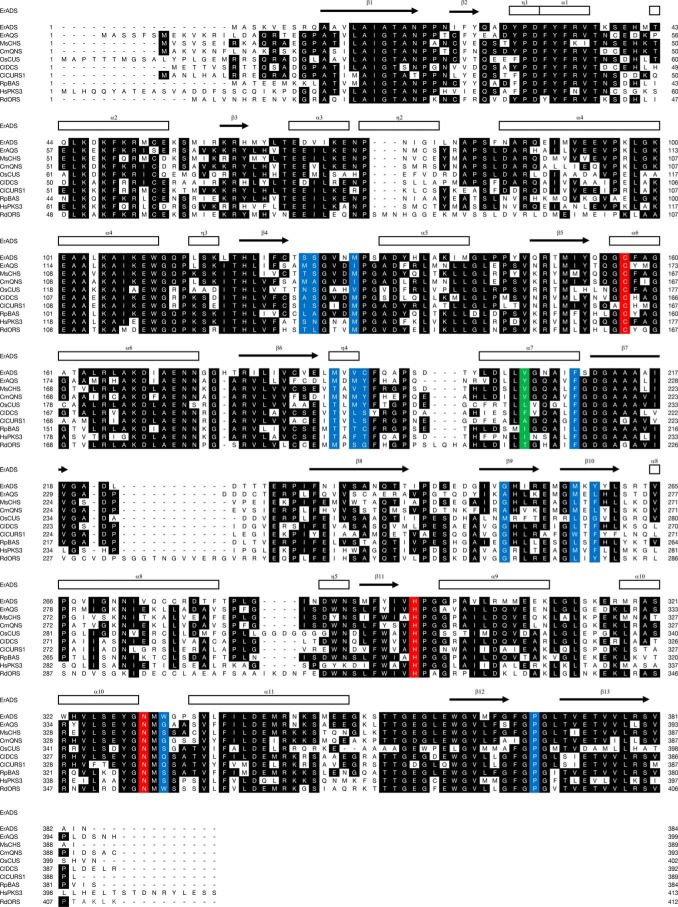
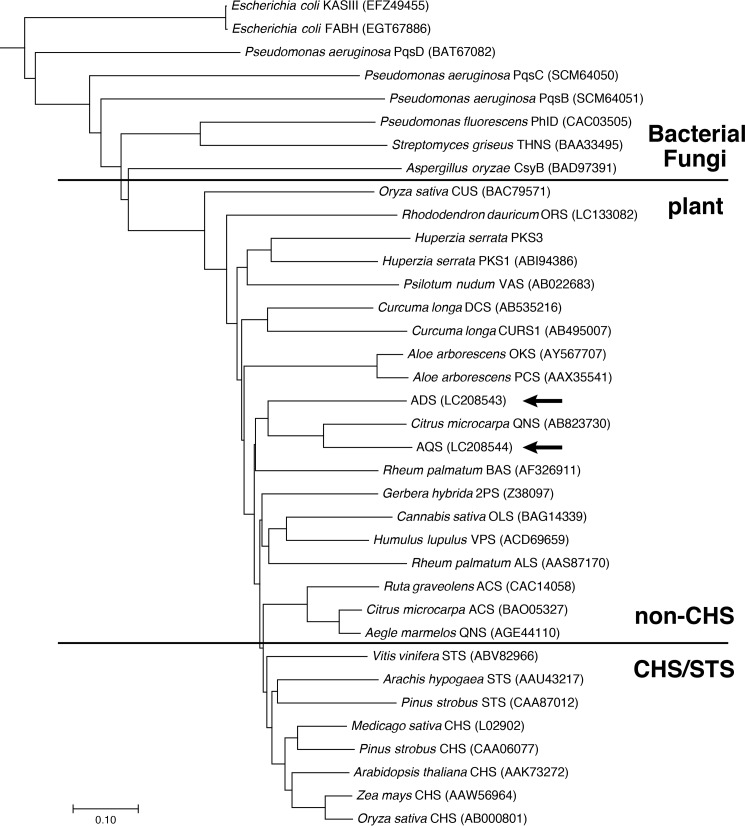
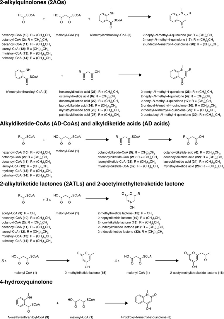
2-Alkylquinolone (2AQ) alkaloids are pharmaceutically and biologically important natural products produced by both bacteria and plants, with a wide range of biological effects, including antibacterial, cytotoxic, anticholinesterase, and quorum-sensing signaling activities. These diverse activities and 2AQ occurrence in vastly different phyla have raised much interest in the biosynthesis pathways leading to their production. Previous studies in plants have suggested that type III polyketide synthases (PKSs) might be involved in 2AQ biosynthesis, but this hypothesis is untested. To this end, we cloned two novel type III PKSs, alkyldiketide-CoA synthase (ADS) and alkylquinolone synthase (AQS), from the 2AQ-producing medicinal plant, Evodia rutaecarpa (Rutaceae). Functional analyses revealed that collaboration of ADS and AQS produces 2AQ via condensations of N-methylanthraniloyl-CoA, a fatty acyl-CoA, with malonyl-CoA. We show that ADS efficiently catalyzes the decarboxylative condensation of malonyl-CoA with a fatty acyl-CoA to produce an alkyldiketide-CoA, whereas AQS specifically catalyzes the decarboxylative condensation of an alkyldiketide acid with N-methylanthraniloyl-CoA to generate the 2AQ scaffold via C-C/C-N bond formations. Remarkably, the ADS and AQS crystal structures at 1.80 and 2.20 Å resolutions, respectively, indicated that the unique active-site architecture with Trp-332 and Cys-191 and the novel CoA-binding tunnel with Tyr-215 principally control the substrate and product specificities of ADS and AQS, respectively. These results provide additional insights into the catalytic versatility of the type III PKSs and their functional and evolutionary implications for 2AQ biosynthesis in plants and bacteria.

Keywords: Evodia rutaecarpa; alkaloid; alkyldiketide synthase; alkylquinolone synthase; biosynthesis; crystal structure; enzyme; polyketide; secondary metabolism; type III polyketide synthase.


Conflict of interest statement

The authors declare that they have no conflicts of interest with the contents of this article

Figures

Figure 1.
Figure 1.
Enzymatic formation of 2AQs and curcuminoids. A, 2-heptyl-4-quinolone formed by PqsB, -C, -D, and -E; B, 2-heptyl-N-methyl-4-quinolone (4) formed by HsPKS3; C, feruloyldiketide-CoA formed by ClDCS; D, curcumin formed by ClCURS1; E, decanoyldiketide-CoA (21) formed by ADS; F, 2-nonyl-N-methyl-4-quinolone (17) formed by AQS.
Figure 2.
Figure 2.
Comparison of the primary sequences of ADS, AQS, and other type III PKSs. The secondary structures of ADS are delineated as follows: α-helices (rectangles) and β-strands (arrows). The Cys, His, and Asn catalytic triad and the residues lining the active-site cavity of CHS are colored red and blue, respectively. Tyr-215 in AQS and the corresponding residues in other type III PKSs are highlighted in green. GenBankTM accession numbers are as follows: MsCHS, L02902; CmQNS, AB823730; ClDCS, AB535216; ClCURS1, AB495007; OsCUS, BAC79571; RpBAS, AF326911; RdORS, LC133082. The sequence of HsPKS3 has not yet been released from GenBankTM.
Figure 3.
Figure 3.
Phylogenetic tree analysis of plant type III PKSs. The bacterial β-ketoacyl carrier protein synthase IIIs (KAS III and FABH) from E. coli were employed as out groups. The scale represents 0.1 amino acid substitutions per site. ADS and AQS are highlighted with arrows. ACS, acridone synthase; ALS, aloesone synthase; BAS, benzalacetone synthase; CURS1, curcumin synthase 1; CUS, curcuminoid synthase; DCS, diketide-CoA synthase; OKS, octaketide synthase; OLS, olivetol synthase; ORS, orcinol synthase; PCS, pentaketide chromone synthase; 2PS, 2-pyrone synthase; STS, stilbene synthase; THNS, tetrahydroxynaphthalene synthase; VAS, phloroisovalerophenone synthase; VPS, valerophenone synthase. GenBankTM registration numbers are shown in parentheses.
Figure 4.
Figure 4.
Substrates used and products obtained in this study.
Figure 5.
Figure 5.
Enzyme reaction products by co-incubation and sole incubations of ADS and AQS. Shown are the enzyme reaction products from N-methylanthraniloyl-CoA (3), malonyl-CoA (1), and fatty acyl-CoAs (hexanoyl (10)-, octanoyl (2)-, decanoyl (11)-, lauroyl (12)-, myristoyl (13)-, or palmitoyl (14)-CoAs) or p-coumaroyl-CoA (7) by the co-incubation of ADS and AQS (A), the sole incubation of ADS (B), and the sole incubation of AQS (C).
Figure 6.
Figure 6.
HPLC elution profiles of EtOAc-soluble enzyme reaction products by the co-incubation of ADS, AQS, and CmQNS and by the sole incubation of CmQNS and AQS. Shown are enzyme reaction products from N-methylanthraniloyl-CoA (3), malonyl-CoA (1), and fatty acyl-CoAs (hexanoyl-CoA (10), octanoyl-CoA (2), decanoyl-CoA (11), lauroyl-CoA (12), or myristoyl-CoA (13)) or p-coumaroyl-CoA (7) by the co-incubation of ADS and CmQNS (A), the co-incubation of AQS and CmQNS (B), and the sole incubation of CmQNS (C). D, enzyme reaction products from 1 and 10, 2, 11-14, 3, or 7 by the sole incubation of AQS.
Figure 7.
Figure 7.
Analysis of all ADS enzyme reaction products. A, the ADS reaction products from decanoyl-CoA (11) and malonyl-CoA (1) at pH 6.5, 7.0, and 7.5. B, analysis of the thioesterase activity of ADS with decanoyldiketide-CoA (21) at pH 6.5, 7.0, and 7.5. The HPLC elution profiles obtained by ADS and boiled ADS are indicated as black and red lines, respectively.
Figure 8.
Figure 8.
Enzyme reaction products from ADS and the ADS mutant enzymes with structurally distinct starter substrates. A–H, the TLC-based analysis of radiolabeled products from malonyl-CoA (1) and hexanoyl-CoA (10) (A), octanoyl-CoA (2) (B), decanoyl-CoA (11) (C), lauroyl-CoA (12) (D), myristyl-CoA (13) (E), palmitoyl-CoA (14) (F), N-methylanthraniloyl-CoA (3) (G), and p-coumaroyl-CoA (7) (H) by the ADS and ADS mutant enzymes. I, HPLC elution profiles of enzyme reaction products obtained by ADS and the boiled ADS reactions with acetyl-CoA (9) and 1 as the substrates.
Figure 9.
Figure 9.
pH-dependent AD acid- and 2ATL-forming activities of ADS. Shown are the relative yields of octanoyldiketide acid (6) and 2-heptyltrikeide lactone (19) from malonyl-CoA (1) and octanoyl-CoA (2) (A), decanoyldiketide acid (22) and 2-nonyltriketide lactone (18) from 1 and decanoyl-CoA (11) (B), and lauroyldiketide acid (24) from 1 and lauroyl-CoA (12) (C). The yields of AD acids at pH 6.0–8.0, 2ATL at pH 5.5 and pH 6.0, and 2ATL at pH 6.0–8.0 are indicated as closed squares and closed and open circles, respectively.
Figure 10.
Figure 10.
Enzyme reaction products from AQS and the AQS Y215V mutant enzyme with structurally distinct substrates. A, enzyme reaction products from N-methylanthraniloyl-CoA (3) as the starter substrate and decanoyldiketide-CoA (21) or decanoyldiketide acid (22) as the extender substrate by AQS and the boiled AQS. B and C, enzyme reaction products from 3 and AD acids (hexanoyl (25)-, octanoyl (6)-, decanoyl (22)-, lauroyl- (24), myristoyl (26)-, or palmitoyl (27)-diketide acid) or p-coumaroyl-CoA (7) by AQS and its boiled enzyme (B) and the AQS Y215V mutant enzyme and its boiled enzyme (C). As the negative control data, the HPLC elution profiles of the enzyme reaction products from 3 and 22 by the boiled enzyme are shown. D, enzyme reaction products from 3 and malonyl-CoA (1) by AQS, the AQS Y215V mutant enzyme, and CmQNS.
Figure 11.
Figure 11.
Overall structures of ADS and AQS. A and B, overall structures of ADS (A) and AQS (B). ADS and AQS form homodimeric structures. The monomers are indicated as blue and gray schematic models, respectively. The CoASH molecule is shown as a green stick model. C–F, close-up views of the active-site entrances of ADS (C), RpBAS (D), MsCHS (E), and the ADS W332G mutant enzyme (F). G, stereo view of the active-site cavity of the docking model of the ADS-bound decanoyl moiety to the catalytic center Cys. The decanoyl moiety is indicated by a pink stick model.
Figure 12.
Figure 12.
Comparison of the active-site architectures of ADS, AQS, their mutant enzymes, and other type III PKSs. Shown are surface representations of ADS (A), RpBAS (B), the ADS W332Q mutant enzyme (C), the ADS W332G mutant enzyme (D), AQS (E), ClCURS1 (F), CmQNS (G), and the AQS Y215V mutant enzyme (H). The enzyme residues and the CoASH molecules are shown as blue and green stick models. The entrances of the CoA-binding tunnels are indicated with arrows. The catalytic triads and the mutated residues are highlighted with red and cyan type, respectively.
Figure 13.
Figure 13.
Analyses of ADS and AQS transcripts and tissue distributions of 2AQs in E. rutaecarpa. A, semiquantitative RT-PCR analyses of ADS and AQS gene expression. The 18S rRNA gene fragment was amplified as a housekeeping gene. B, 2AQs contents in leaves, buds, and fruits, analyzed by LC-ESI-MS. The data are presented as the mean ± S.D. (n = 3). The yields of the C7, C9, C11, and C13 2AQs 4, 17, 20, and 29 are represented as white, gray, black, and dotted bars, respectively. The amounts of 2AQs were estimated from the peak intensities, by comparison with those of the corresponding authentic 2AQs, and then converted to the yield percentage against the wet weights of the tissues, respectively.

References

    1. Wang X. X., Zan K., Shi S. P., Zeng K. W., Jiang Y., Guan Y., Xiao C. L., Gao H. Y., Wu L. J., and Tu P. F. (2013) Quinolone alkaloids with antibacterial and cytotoxic activities from the fruits of Evodia rutaecarpa. Fitoterapia 89, 1–7 - PubMed
    1. Cardoso-Lopes E. M., Maier J. A., da Silva M. R., Regasini L. O., Simote S. Y., Lopes N. P., Pirani J. R., Bolzani Vda S., and Young M. C. (2010) Alkaloids from stems of Esenbeckia leiocarpa Engl. (Rutaceae) as potential treatment for Alzheimer disease. Molecules 15, 9205–9213 - PMC - PubMed
    1. Heeb S., Fletcher M. P., Chhabra S. R., Diggle S. P., Williams P., and Cámara M. (2011) Quinolones: from antibiotics to autoinducers. FEMS Microbiol. Rev. 35, 247–274 - PMC - PubMed
    1. Wang C. F., Fan L., Tian M., Qi X. S., Liu J. X., Feng J. B., Du S. S., Su X., and Wang Y. Y. (2014) Radiosensitizing effect of schinifoline from Zanthoxylum schinifolium Sieb et Zucc on human non-small cell lung cancer A549 cells: a preliminary in vitro investigation. Molecules 19, 20128–20138 - PMC - PubMed
    1. Dulcey C. E., Dekimpe V., Fauvelle D. A., Milot S., Groleau M. C., Doucet N., Rahme L. G., Lépine F., and Déziel E. (2013) The end of an old hypothesis: the Pseudomonas signaling molecules 4-hydroxy-2-alkylquinolines derive from fatty acids, not 3-ketofatty acids. Chem. Biol. 20, 1481–1491 - PMC - PubMed

Publication types