. 2017 Dec 1;432(1):72-85.
doi: 10.1016/j.ydbio.2017.04.002. Epub 2017 Apr 12.

Spop regulates Gli3 activity and Shh signaling in dorsoventral patterning of the mouse spinal cord

Affiliations

Spop regulates Gli3 activity and Shh signaling in dorsoventral patterning of the mouse spinal cord

Hongchen Cai et al. Dev Biol. .

Abstract

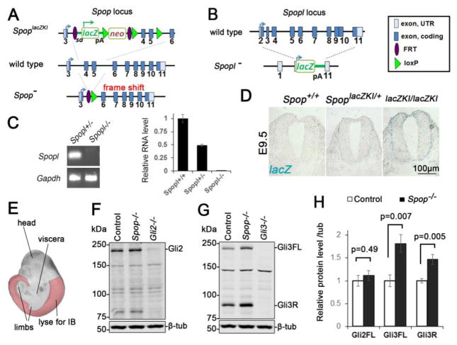
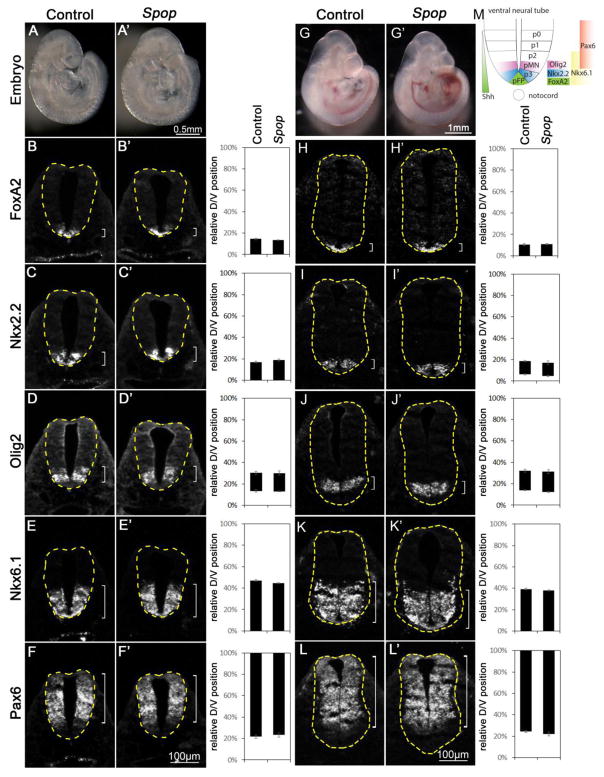
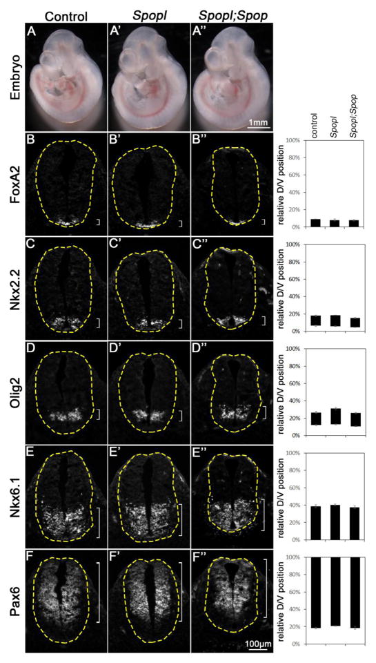
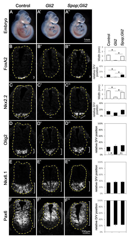
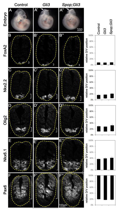
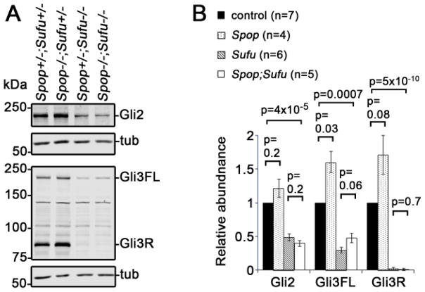
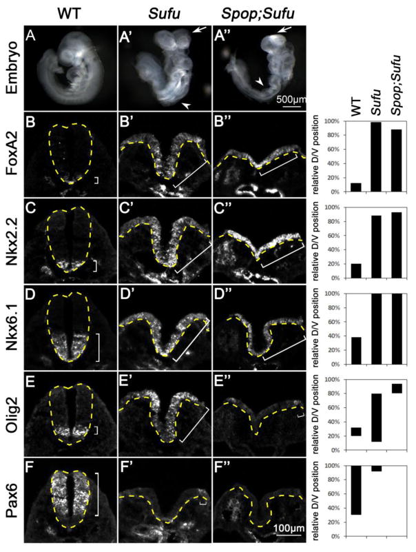
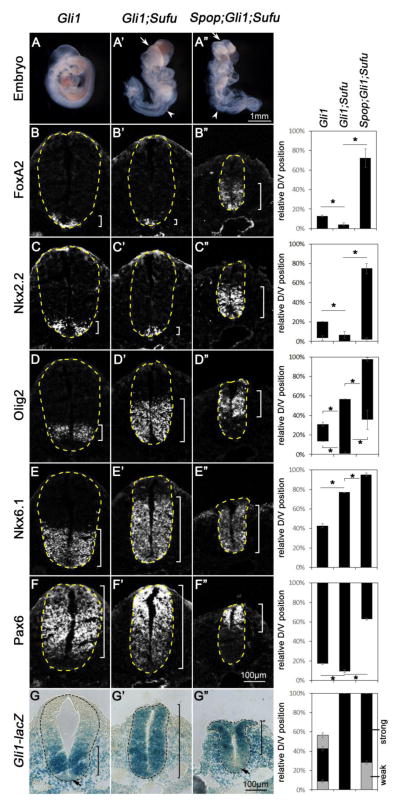
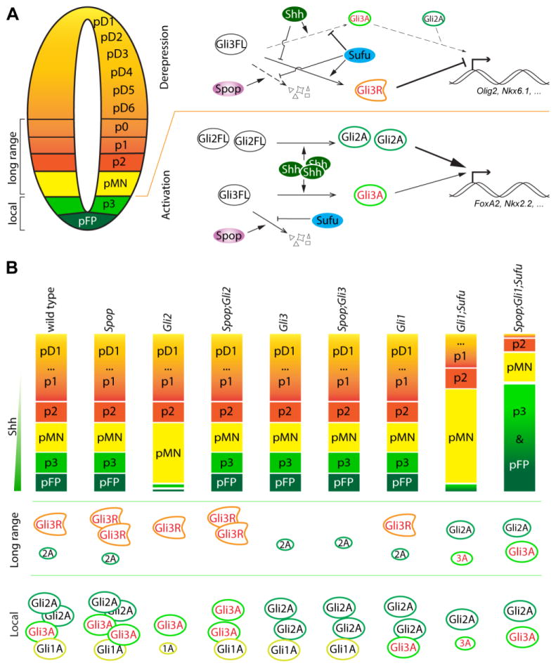
Sonic Hedgehog (Shh) signaling regulates the patterning of ventral spinal cord through the effector Gli family of transcription factors. Previous in vitro studies showed that an E3 ubiquitin ligase containing Speckle-type POZ protein (Spop) targets Gli2 and Gli3 for ubiquitination and degradation, but the role of Spop in Shh signaling and mammalian spinal cord patterning remains unknown. Here, we show that loss of Spop does not alter spinal cord patterning, but it suppresses the loss of floor plate and V3 interneuron phenotype of Gli2 mutants, suggesting a negative role of Spop in Gli3 activator activity, Shh signaling and the specification of ventral cell fates in the spinal cord. This correlates with a moderate but significant increase in the level of Gli3 protein in the Spop mutant spinal cords. Furthermore, loss of Spop restores the maximal Shh pathway activation and ventral cell fate specification in the Gli1;Sufu double mutant spinal cord. Finally, we show that loss of Spop-like does not change the spinal cord patterning in either wild type or Spop mutants, suggesting that it does not compensate for the loss of Spop in Shh signaling and spinal cord patterning. Therefore, our results demonstrate a negative role of Spop in the level and activity of Gli3, Shh signaling and ventral spinal cord patterning.

Keywords: Gli1; Gli2; Neural patterning; Spopl; Sufu; Ubiquitin ligase.


Figures

Fig. 1
Fig. 1. A moderate increase in Gli3 protein level in the Spop mutant spinal cords
(A) A schematic illustration of Spop loss-of-function mutant alleles. SpoplacZKI contained a lacZ and neomycin resistance cassette. SpopΔEx was generated from recombination of introduced FRT and loxP sites that deletes the 4th and 5th exons. This deletion also resulted in a frame shift that truncated the protein. (B) A schematic illustration of Spopl null mutant allele, in which a lacZ reporter replaced the entire protein-coding region of Spopl. pA: polyA signal. (C) Quantitative real time PCR showing the absence of Spopl transcript in E9.5 Spopl mutant embryos (mean ± SEM, n=3 wild type, 4 Spopl+/− and 5 Spopl−/− embryos). (D) X-gal staining of E9.5 SpoplacZKI heterozygotes and homozygotes showing Spop expression in the neural tube. (E) A schematic illustration of tissue (highlighted region) used for immunoblot in (F) and (G). The trunks of E10.5 embryos were freed of viscera and limbs to minimize the effect of Gli proteins in these tissues. (F) Immunoblots with antibodies against Gli2 and β-tubulin. (G) Immunoblots with antibodies against Gli3 and β-tubulin. (H) Quantification of (F) and (G) (mean ± SEM from n=6 embryos per group). Student’s t-test showed a significant increase in the levels of Gli3FL and Gli3R, but not Gli2 in Spop mutants.
Fig. 2
Fig. 2. Normal ventral patterning of the Spop mutant spinal cords
(A, A′) Lateral views of E9.5 control (A) and Spop (A′) mutant embryos. (B–F′) Immunofluorescent images of transverse sections of E9.5 control (n=4 embryos) and Spop mutant (n=4 embryos) spinal cords at the forelimb level with indicated antibodies. (G, G′) Lateral views of E10.5 control (G) and Spop (G′) mutant embryos. (H–L′) Immunofluorescent images of transverse sections of E10.5 control (n=4 embryos) and Spop mutant (n=4 embryos) spinal cords at the forelimb level. The spinal cords are outlined with dash lines. Brackets indicate the expression domains. The span of each domain along the D/V axis is quantified and shown on the right. Relative D/V position is shown as the distance to the ventral-most point of the spinal cord as a percentage of the entire D/V span of the spinal cord. Student’s t-test suggests no significant difference between Spop mutant and control groups. (M) A schematic illustration of ventral spinal cord patterning at E10.5 depicting the arrangement of various progenitor groups and the expression domains of marker genes in wild type embryos.
Fig. 3
Fig. 3. Normal ventral patterning of the Spopl mutant and Spopl;Spop double mutant spinal cords
(A–A″) Lateral views of E10.5 control (A), Spop mutant (A′) and Spopl;Spop double mutant (A″) embryos. (B–F″) Immunofluorescent images of transverse sections of E10.5 control (n=4 embryos), Spopl mutant (n=4 embryos) and Spopl;Spop double mutant (n=4 embryos) spinal cords at the forelimb level with indicated antibodies are shown. The spinal cords are outlined with dash lines. Brackets indicate the expression domains. The span of each domain along the D/V axis is quantified and shown on the right. Relative D/V position is shown as the distance to the ventral-most point of the spinal cord as a percentage of the entire D/V span of the spinal cord. Student’s t-test suggests no significant difference between Spopl;Spop double mutant, Spopl mutant and control groups. The morphology and spinal cord neural patterning of both Spopl mutant and Spopl;Spop double mutant resemble wild type.
Fig. 4
Fig. 4. Restored floor plate and V3 interneuron progenitor fates in Spop;Gli2 double mutants
(A–A″) Lateral views of E10.5 control (A), Gli2 mutant (A′) and Spop;Gli2 double mutant (A″) embryos. (B–F″) Immunofluorescent images of transverse sections of E10.5 control (n=3 embryos), Gli2 mutant (n=3 embryos) and Spop;Gli2 (n=3 embryos) double mutant spinal cords at the forelimb level with indicated antibodies. The spinal cords are outlined with dash lines. Brackets indicate the expression domains. The span of each domain along D/V axis is shown on the right. Relative D/V position is shown as the distance to the ventral-most point of the spinal cord as a percentage of the entire D/V span of the spinal cord. The width of Foxa2 and Nkx2.2 domains is also quantified (hollow columns). (B–B″) Foxa2 was present in the ventral-most region of the control and diminished in the Gli2 mutant, but restored in the Spop;Gli2 double mutant spinal cords. Student’s t-test was employed to compare the width of floor plate. *: p<0.05. (C–C″) Nkx2.2 was present in juxtaposition to floor plate of the control but diminished in the Gli2 mutant, and was restored in the Spop;Gli2 double mutant spinal cords. Student’s t-test was employed to compare the size of V3 interneuron progenitor domain. *: p<0.05. (D–D″) Olig2 expression was excluded from the ventral-most region of control and Spop;Gli2 double mutant but expanded ventrally in Gli2 mutant spinal cords. Student’s t-test was employed to compare the distance from the Olig2 expression domain to the ventral-most point of the spinal cords. *: p<0.05. (E–F″) Nkx6.1 and Pax6 expression domains remained unchanged in the Gli2 mutant and Spop;Gli2 double mutant spinal cords.
Fig. 5
Fig. 5. Spop;Gli3 double mutants resembled Gli3 mutants in ventral spinal cord patterning
(A–A″) Lateral views of E10.5 control (A), Gli3 mutant (A′) and Spop;Gli3 double mutant (A″) embryos. Arrows in A′ and A″ indicate exencephaly. (B–F″) Immunofluorescent images of transverse sections of E10.5 spinal cords in Gli3 mutant (n=4 embryos) and Spop;Gli3 double mutant (n=5 embryos) resembled those in the control (n=5 embryos) at the forelimb level with indicated antibodies. The spinal cords are outlined with dash lines. Brackets indicate the expression domains. The span of each domain is quantified and shown on the right. Relative D/V position is shown as the distance to the ventral-most point of the spinal cord as a percentage of the entire D/V span of the spinal cord. Student’s t-test suggests no significant difference between Spop;Gli3 double mutant, Gli3 mutant and control groups.
Fig. 6
Fig. 6. Moderate increase in the level of Gli3, but not Gli2, in Spop;Sufu double mutant embryos
(A) Immunoblots of E9.5 embryo lysates with indicated antibodies. (B) Quantitative analyses of the levels of Gli2, Gli3FL and Gli3R (normalized to β-tubulin, mean ± SEM) based on the data of immunoblot analyses. Statistical significance was determined with Student’s t-test.
Fig. 7
Fig. 7. Loss of Spop exacerbated the ventralization of the Sufu mutant spinal cord
(A–C) Lateral views of E9.5 wild type (A), Sufu mutant (A′) and Spop;Sufu double mutant (A″) embryos. Arrows in B and C indicate exencephaly; arrowheads indicate spina bifida. (B–F″) Immunofluorescent images of transverse sections of E9.5 spinal cords at the thoracic level. The spinal cords are outlined with dash lines. Brackets indicate the expression domains. (B–C″) Foxa2 and Nkx2.2 were expressed in the ventral-most region of wild type but expanded to the dorsal region in the Sufu mutant and Spop;Sufu double mutant spinal cords. (D–D″) Nkx6.1 expression domain was expanded to the dorsal region in both Sufu mutant and Spop;Sufu double mutant spinal cords. (E–E″) Olig2 expression was shifted to the dorsal region in Sufu mutant and even more dorsally shifted in Spop;Sufu double mutant spinal cords. (F–F″) Pax6 expression domain was shifted dorsally in Sufu mutant and was absent in Spop;Sufu double mutant spinal cord. n=2 embryos for each genotype were analyzed. The span of each domain is quantified and shown on the right. Relative D/V position is shown as the distance to the ventral-most point of the spinal cord as a percentage of the entire D/V span of the spinal cord.
Fig. 8
Fig. 8. Loss of Spop rescued the floor plate and V3 interneuron progenitor fates in Gli1;Sufu double mutant embryos
(A–A″) Lateral views of E9.5 Gli1 mutant (A), Gli1;Sufu double mutant (A′) and Spop;Gli1;Sufu triple mutant (A″) embryos. Arrows in A′ and A″ indicate exencephaly; white arrowheads indicate spina bifida; red arrowheads indicate edema. (B–G″) Immunofluorescent (B–F″) or Xgal-stained (G–G″) images of transverse sections of the E9.5 Gli1 mutant (n=3 embryos), Gli1;Sufu double mutant (n=3 embryos) and Spop;Gli1;Sufu triple mutant (n=3 embryos) spinal cords at the thoracic level. The spinal cords are outlined with dash lines. Brackets indicate the expression domains. The span of each domain along the D/V axis is shown on the right. Relative D/V position is shown as the distance to the ventral-most point of the spinal cord as a percentage of the entire D/V span of the spinal cord. Student’s t-tests were performed to compare the dorsal and ventral borders of expression domains of various genes. *: p<0.05. (B–C″) The expression of Foxa2 and Nkx2.2 in the ventral-most region of Gli1 mutant was diminished in Gli1;Sufu double mutant, but expanded dorsally in the Spop;Gli1;Sufu triple mutant spinal cords. (D–D″) Olig2 expression was expanded both ventrally and dorsally in Gli1;Sufu double mutant, but was dorsally restricted in the Spop;Gli1;Sufu triple mutant spinal cords. (E–E″) Nkx6.1 expression domain was expanded dorsally in the Gli1;Sufu double mutant and more dorsally in the Spop;Gli1;Sufu triple mutant spinal cords. (F–F″) Pax6 expression domain was expanded ventrally in the Gli1;Sufu double mutant, and restricted to the more dorsal region in the Spop;Gli1;Sufu triple mutant spinal cord. (G) Gli1-lacZ expression formed a ventral-to-dorsal gradient in the Gli1 mutant spinal cord, with the exception of the floor plate, in which Gli1-lacZ has been downregulated. (G′) Strong Gli1-lacZ expression is present throughout the entire spinal cord of Gli1;Sufu double mutant. (G″) Gli1-lacZ is highly expressed in the dorsal, but not ventral region of the Spop;Gli1;Sufu triple mutant spinal cords.
Fig. 9
Fig. 9. A model of the roles of Spop in spinal cord patterning
(A) In the lateral region of the spinal cord (long range) including p0-pMN, low to intermediate levels of Shh reduce the production of Gli3R from Gli3FL by antagonizing the repressive function of Sufu, allowing the expression of genes such as Olig2 and Nkx6.1. In pFP and p3, high levels of Shh promote the production of Gli2A and Gli3A, which then activate the expression of Foxa2 and Nkx2.2. Gli2A plays a more predominant role than Gli3A in this context. Spop targets Gli3 for degradation, preventing over activation of the Shh pathway. (B) The levels of both Gli3A and Gli3R increase in the absence of Spop, but the effect of the increased Gli3A on the formation of pFP and p3 is only revealed in Spop;Gli2 double mutants, in which the much more potent Gli2A is absent. In Gli1;Sufu double mutants, the reduced levels of Gli2A and Gli3A are insufficient to support pFP and p3 formation. Loss of Spop increases Gli3A and rescues the formation of pFP and p3 in Sufu;Gli1;Spop triple mutants. The moderate increase in Gli3R in the absence of Spop does not show apparent effect in ventral spinal cord patterning in various single and compound mutants.

Similar articles

Cited by

References

    1. Bai CB, Auerbach W, Lee JS, Stephen D, Joyner AL. Gli2, but not Gli1, is required for initial Shh signaling and ectopic activation of the Shh pathway. Development. 2002;129:4753–61. - PubMed
    1. Bai CB, Joyner AL. Gli1 can rescue the in vivo function of Gli2. Development. 2001;128:5161–72. - PubMed
    1. Bai CB, Stephen D, Joyner AL. All mouse ventral spinal cord patterning by hedgehog is Gli dependent and involves an activator function of Gli3. Dev Cell. 2004;6:103–15. - PubMed
    1. Balaskas N, Ribeiro A, Panovska J, Dessaud E, Sasai N, Page KM, Briscoe J, Ribes V. Gene regulatory logic for reading the Sonic Hedgehog signaling gradient in the vertebrate neural tube. Cell. 2012;148:273–84. - PMC - PubMed
    1. Barnfield PC, Zhang X, Thanabalasingham V, Yoshida M, Hui CC. Negative regulation of Gli1 and Gli2 activator function by Suppressor of fused through multiple mechanisms. Differentiation. 2005;73:397–405. - PubMed

MeSH terms

LinkOut - more resources