. 2017 Apr 15;6(4):489-495.
doi: 10.1242/bio.024133.

Tanshinone IIA suppresses the progression of atherosclerosis by inhibiting the apoptosis of vascular smooth muscle cells and the proliferation and migration of macrophages induced by ox-LDL

Affiliations

Tanshinone IIA suppresses the progression of atherosclerosis by inhibiting the apoptosis of vascular smooth muscle cells and the proliferation and migration of macrophages induced by ox-LDL

Baocai Wang et al. Biol Open. .

Expression of concern in

Abstract

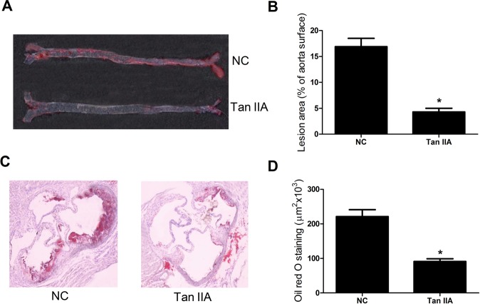
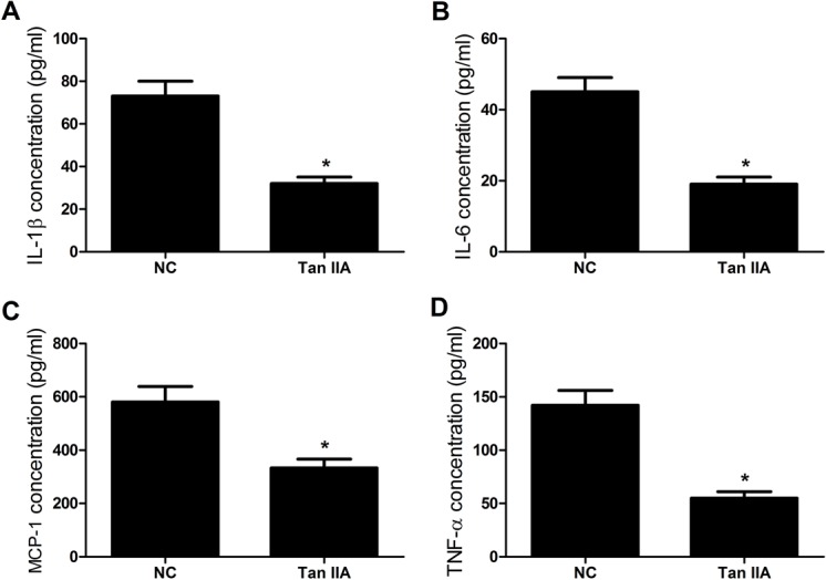
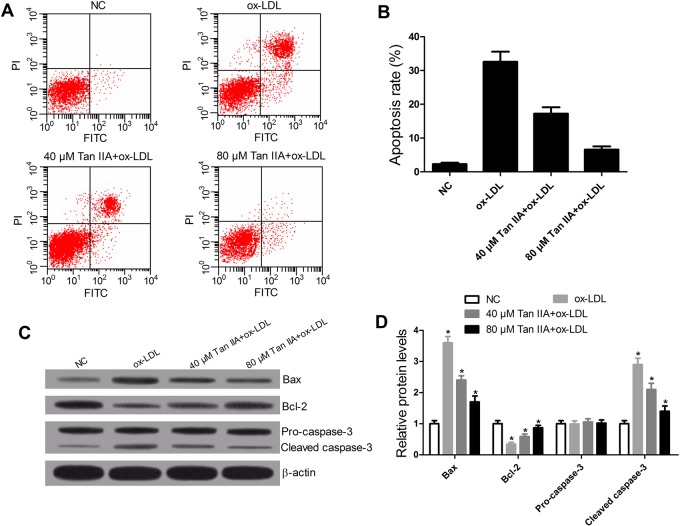
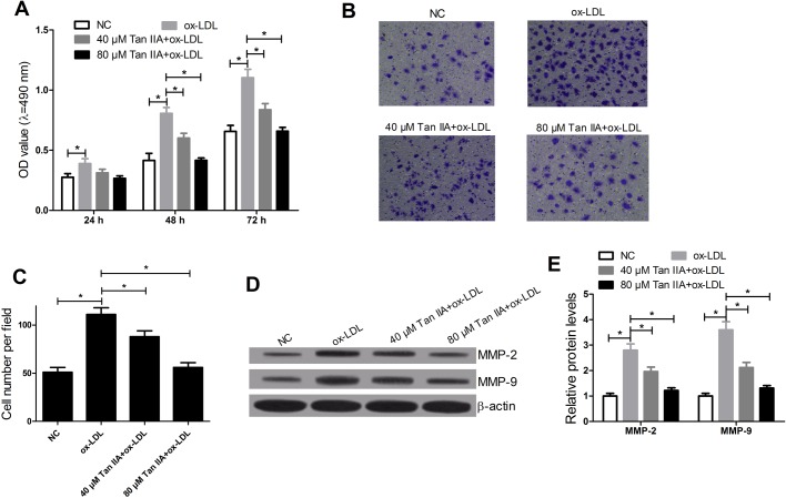
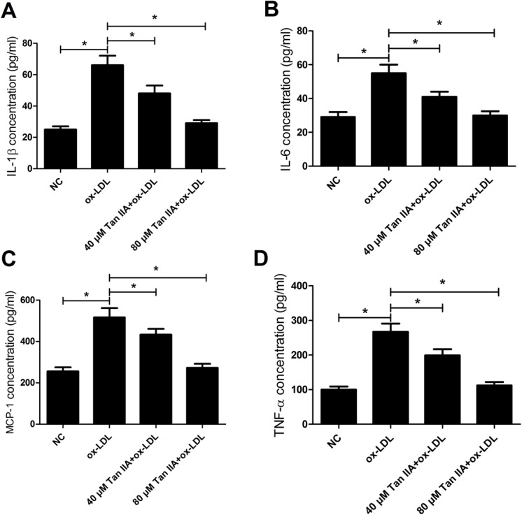
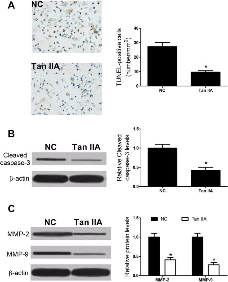
The profound inhibitory effect of Tanshinone IIA (Tan IIA) on atherosclerosis has been demonstrated, whereas the latent mechanism is not completely cleared. This study aimed to investigate the cellular and molecular mechanisms underlying Tan IIA protecting against atherosclerosis. Oil Red O staining and ELISA assay showed that Tan IIA suppressed the progress of atherosclerosis and reduced the levels of inflammatory cytokines in serum of apolipoprotein E deficient (ApoE -/- ) mice. Flow cytometry assay revealed that Tan IIA inhibited oxidized LDL (ox-LDL)-induced apoptosis of VSMCs. MTT and transwell assay indicated that Tan IIA suppressed the ox-LDL-stimulated proliferation and migration of RAW264.7 cells. Moreover, Tan IIA ameliorated inflammatory cytokine upregulation elicited by ox-LDL in RAW264.7 cells. Additionally, Tan IIA inhibited the apoptosis of VSMCs and decreased the levels of MMP-2, MMP-9 in ApoE-/- mice. In conclusion, our study demonstrated Tan IIA suppressed the progression of atherosclerosis by inhibiting vascular inflammation, apoptosis of VSMCs and proliferation and migration of macrophages induced by ox-LDL.

Keywords: Atherosclerosis; Macrophages; Tanshinone IIA; Vascular smooth muscle cells; ox-LDL.


Conflict of interest statement

Competing interests

The authors declare no competing or financial interests.

Figures

Fig. 1.
Fig. 1.
Effects of Tan IIA on atherosclerosis in ApoE−/− mice. ApoE−/− mice at 6 weeks were gavaged with Tan IIA (30 mg/kg) for 20 weeks and then sacrificed. (A) Representative photographs indicating the atherosclerotic plaque in aortas from ApoE–/– mice with Oil Red O. (B) The atherosclerotic lesion area in the Tan IIA group and control group. (C) Representative photomicrographs of the aortic root from ApoE−/− mice stained with Oil Red O. (D) The atherosclerotic lesion was quantified as the fraction area in Tan IIA group and control group. In B and D, n=8, *P<0.05, error bars indicate that data were expressed as the mean±s.d.
Fig. 2.
Fig. 2.
Tan IIA suppresses expression of inflammatory cytokines in serum of ApoE–/– mice. ApoE−/− mice at 6 weeks were gavaged with Tan IIA (30 mg/kg) for 20 weeks and then sacrificed. (A-D) The concentrations of IL-1β, IL-6, MCP-1, and TNF-α in serum of ApoE–/– mice treated with Tan IIA were significantly decreased by ELISA assay. n=8, *P<0.05, error bars indicate that data were expressed as the mean±s.d.
Fig. 3.
Fig. 3.
Tan IIA inhibits ox-LDL-induced apoptosis of VSMCs through regulating the apoptosis-related protein levels. VSMCs were treated with ox-LDL (50 μg/ml) or ox-LDL+Tan IIA (40 or 80 μM) for 24 h. (A,B) Flow cytometry showed that Tan IIA reversed ox-LDL-induced apoptosis in VSMCs. (C,D) Western blot displayed that Tan IIA overturned the effects of ox-LDL on the expression of Bcl-2, Bax and cleaved caspase-3. n=4, *P<0.05, error bars in B and D indicate that data were expressed as the mean±s.d.
Fig. 4.
Fig. 4.
Tan IIA inhibited the ox-LDL-induced proliferation and migration of RAW264.7 cells. RAW264.7 cells were treated with ox-LDL (50 μg/ml) or Tan IIA (40 or 80 μM). (A) MTT was used to measure the effect of ox-LDL and Tan IIA on cell proliferation. (B,C) Transwell chamber was performed to detect the effect of ox-LDL and Tan IIA on cell migration after treatment with ox-LDL or (ox-LDL+Tan IIA) for 24 h. (D,E) Western blots were carried out to determine the levels of MMP-9 and MMP-2 after treatment with ox-LDL or (ox-LDL+Tan IIA) for 24 h in RAW264.7 cells. n=4, *P<0.05, error bars in A, C and E indicate that data were expressed as the mean±s.d.
Fig. 5.
Fig. 5.
Tan IIA ameliorates inflammatory cytokine upregulation in ox-LDL-stimulated RAW264.7 cells. RAW264.7 cells were treated with ox-LDL (50 μg/ml) or (ox-LDL+Tan IIA) (40 or 80 μM) for 24 h. (A-D) The concentrations of inflammatory cytokines (IL-1β, IL-6, MCP-1, and TNF-α) in RAW264.7 cells treated with ox-LDL or (ox-LDL+Tan IIA) were detected by ELISA. n=4, *P<0.05, error bars indicate that data were expressed as the mean±s.d.
Fig. 6.
Fig. 6.
Tan IIA reduced the apoptosis rate of VSMCs and decreased the levels of MMP-2 and MMP-9 in ApoE−/− mice. (A) TUNEL assay showing decrease in TUNEL-positive cells in ApoE−/− mice treated with Tan IIA against to control group. (B,C) Western blots confirming the levels of cleaved caspase-3 or that the migration-related proteins MMP-2 and MMP-9 decreased when treated with Tan IIA in ApoE−/− mice, *P<0.05, error bars indicate that data were expressed as the mean±s.d. (n=8).

Similar articles

Cited by

References

    1. Andrés V., Pello O. M. and Silvestre-Roig C. (2012). Macrophage proliferation and apoptosis in atherosclerosis. Curr. Opin. Lipidol. 23, 429-438. 10.1097/MOL.0b013e328357a379 - DOI - PubMed
    1. Chen Z. and Xu H. (2014). Anti-inflammatory and immunomodulatory mechanism of tanshinone IIA for atherosclerosis. Evid. Based. Complement. Alternat. Med. 2014, 267976 10.1155/2014/267976 - DOI - PMC - PubMed
    1. Chistiakov D. A., Orekhov A. N. and Bobryshev Y. V. (2015). Vascular smooth muscle cell in atherosclerosis. Acta. Physiol. 214, 33-50. 10.1111/apha.12466 - DOI - PubMed
    1. Clarke M. C., Figg N., Maguire J. J., Davenport A. P., Goddard M., Littlewood T. D. and Bennett M. R. (2006). Apoptosis of vascular smooth muscle cells induces features of plaque vulnerability in atherosclerosis. Nat. Med. 12, 1075-1080. 10.1038/nm1459 - DOI - PubMed
    1. Cutini P. H. and Massheimer V. L. (2010). Role of progesterone on the regulation of vascular muscle cells proliferation, migration and apoptosis. Steroids 75, 355-361. 10.1016/j.steroids.2010.01.017 - DOI - PubMed

LinkOut - more resources