

Increased reactive oxygen species levels cause ER stress and cytotoxicity in andrographolide treated colon cancer cells
- PMID: 28412728
- PMCID: PMC5432246
- DOI: 10.18632/oncotarget.15393
Increased reactive oxygen species levels cause ER stress and cytotoxicity in andrographolide treated colon cancer cells
Abstract
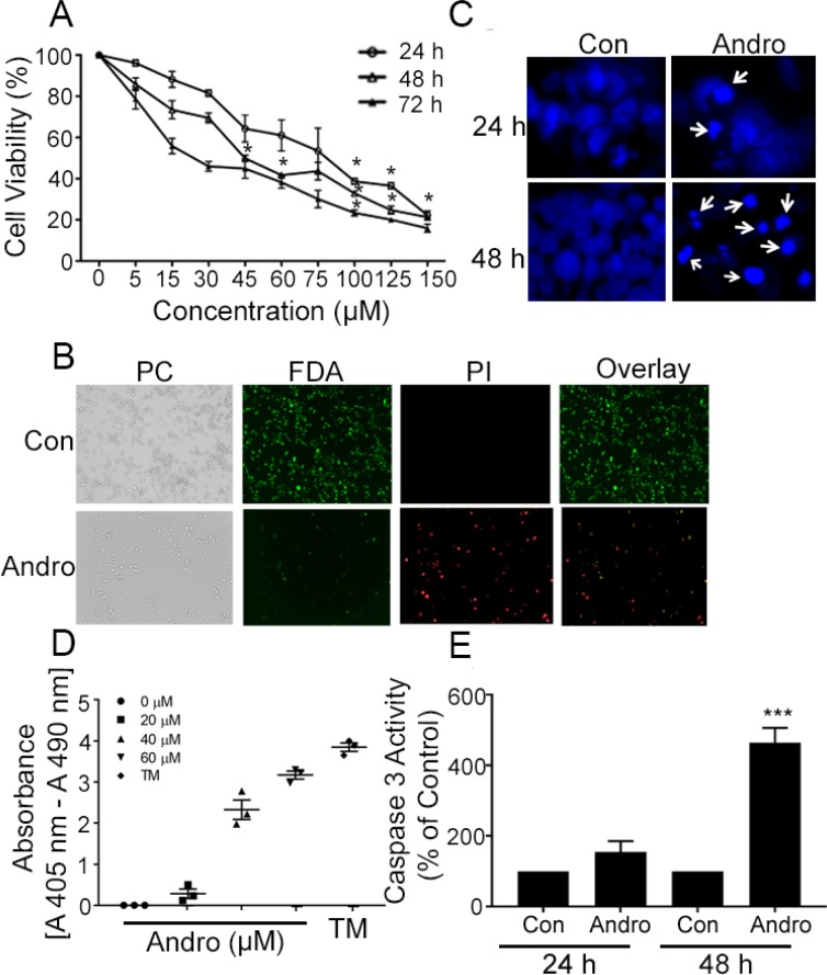
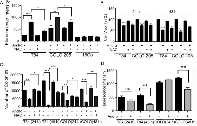
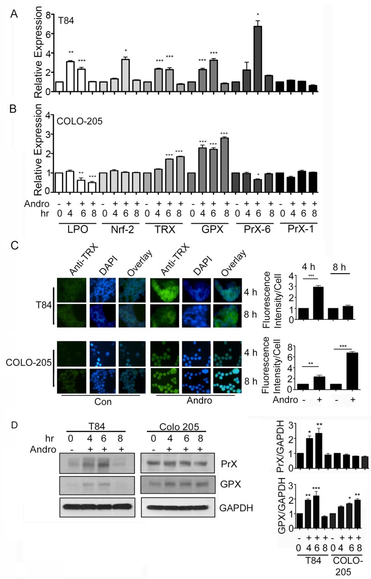
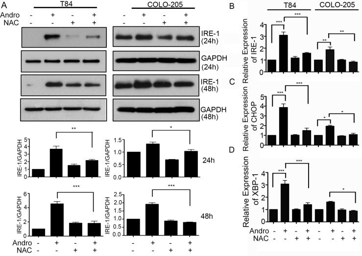
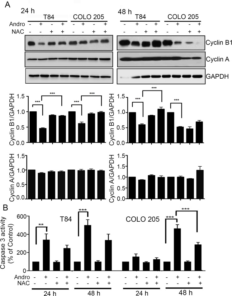
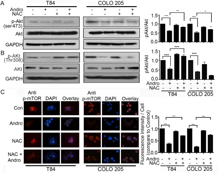
Chemotherapy continues to play an essential role in the management of many cancers including colon cancer, the third leading cause of death due to cancer in the United States. Many naturally occurring plant compounds have been demonstrated to possess anti-cancer cell activity and have the potential to supplement existing chemotherapy strategies. The plant metabolite andrographolide induces cell death in cancer cells and apoptosis is dependent upon the induction of endoplasmic reticulum stress (ER stress) leading to the unfolded protein response (UPR). The goal of the present study was to determine the mechanism by which andrographolide induces ER stress and to further evaluate its role in promoting cell death pathways. The T84 and COLO 205 cancer cell lines were used to demonstrate that andrographolide induces increased ROS levels, corresponding anti-oxidant response molecules, and reduced mitochondrial membrane potential. No increases in ROS levels were detected in control colon fibroblast cells. Andrographolide-induced cell death, UPR signaling, and CHOP, Bax, and caspase 3 apoptosis elements were all inhibited in the presence of the ROS scavenger NAC. Additionally, andrographolide-induced suppression of cyclins B1 and D1 were also reversed in the presence of NAC. Finally, Akt phosphorylation and phospho-mTOR levels that are normally suppressed by andrographolide were also expressed at normal levels in the absence of ROS. These data demonstrate that andrographolide induces ER stress leading to apoptosis through the induction of ROS and that elevated ROS also play an important role in down-regulating cell cycle progression and cell survival pathways as well.
Keywords: andrographolide; chemotherapy; endoplasmic reticulum stress; reactive oxygen species; unfolded protein response.
Conflict of interest statement
None of the authors participating in this study have any conflicts of interest in this work or publication.
Figures
Similar articles
-
Endoplasmic reticulum stress and IRE-1 signaling cause apoptosis in colon cancer cells in response to andrographolide treatment.Oncotarget. 2016 Jul 5;7(27):41432-41444. doi: 10.18632/oncotarget.9180. Oncotarget. 2016. PMID: 27166181 Free PMC article.
-
Chrysin induces death of prostate cancer cells by inducing ROS and ER stress.J Cell Physiol. 2017 Dec;232(12):3786-3797. doi: 10.1002/jcp.25861. Epub 2017 May 3. J Cell Physiol. 2017. PMID: 28213961
-
Curcumin induces ER stress-mediated apoptosis through selective generation of reactive oxygen species in cervical cancer cells.Mol Carcinog. 2016 May;55(5):918-28. doi: 10.1002/mc.22332. Epub 2015 May 15. Mol Carcinog. 2016. PMID: 25980682
-
An Overview of Unfolded Protein Response Signaling and Its Role in Cancer.Cancer Biother Radiopharm. 2017 Oct;32(8):275-281. doi: 10.1089/cbr.2017.2309. Cancer Biother Radiopharm. 2017. PMID: 29053418 Review.
-
Natural antioxidants' effects on endoplasmic reticulum stress-related diseases.Food Chem Toxicol. 2020 Apr;138:111229. doi: 10.1016/j.fct.2020.111229. Epub 2020 Feb 24. Food Chem Toxicol. 2020. PMID: 32105807 Review.
Cited by
-
Cytotoxic Mechanism of Sphaerodactylomelol, an Uncommon Bromoditerpene Isolated from Sphaerococcus coronopifolius.Molecules. 2021 Mar 4;26(5):1374. doi: 10.3390/molecules26051374. Molecules. 2021. PMID: 33806445 Free PMC article.
-
Cytotoxic Effect of Andrographis paniculata Associated with 2-Aminoethyl Dihydrogen Phosphate in Triple-Negative Breast Cells.Curr Issues Mol Biol. 2024 Jan 5;46(1):527-541. doi: 10.3390/cimb46010034. Curr Issues Mol Biol. 2024. PMID: 38248336 Free PMC article.
-
Ablation of catalase promotes non-alcoholic fatty liver via oxidative stress and mitochondrial dysfunction in diet-induced obese mice.Pflugers Arch. 2019 Jun;471(6):829-843. doi: 10.1007/s00424-018-02250-3. Epub 2019 Jan 7. Pflugers Arch. 2019. PMID: 30617744
-
The Role of the ER-Induced UPR Pathway and the Efficacy of Its Inhibitors and Inducers in the Inhibition of Tumor Progression.Oxid Med Cell Longev. 2019 Feb 3;2019:5729710. doi: 10.1155/2019/5729710. eCollection 2019. Oxid Med Cell Longev. 2019. PMID: 30863482 Free PMC article. Review.
-
The Effect of Terpenoid Natural Chinese Medicine Molecular Compound on Lung Cancer Treatment.Evid Based Complement Alternat Med. 2021 Dec 16;2021:3730963. doi: 10.1155/2021/3730963. eCollection 2021. Evid Based Complement Alternat Med. 2021. PMID: 34956377 Free PMC article. Review.
References
-
- World Health . Organization. Herba andrographolis. In: World Health Orgaonization, editor. WHO Monographs on selected medicinal plants. Geneva: World Health Organization; 2002.
-
- Lim JC, Chan TK, Ng DS, Sagineedu SR, Stanslas J, Wong WS. Andrographolide and its analogues: versatile bioactive molecules for combating inflammation and cancer. Clin Exp Pharmacol Physiol. 2012;39:300–310. - PubMed
-
- Lee YC, Lin HH, Hsu CH, Wang CJ, Chiang TA, Chen JH. Inhibitory effects of andrographolide on migration and invasion in human non-small cell lung cancer A549 cells via down-regulation of PI3K/Akt signaling pathway. Eur J Pharmacol. 2010;632:23–32. - PubMed
MeSH terms
Substances
LinkOut - more resources
Full Text Sources
Other Literature Sources
Research Materials
Miscellaneous
