

Germline Mutations in CDH23, Encoding Cadherin-Related 23, Are Associated with Both Familial and Sporadic Pituitary Adenomas
- PMID: 28413019
- PMCID: PMC5420349
- DOI: 10.1016/j.ajhg.2017.03.011
Germline Mutations in CDH23, Encoding Cadherin-Related 23, Are Associated with Both Familial and Sporadic Pituitary Adenomas
Abstract
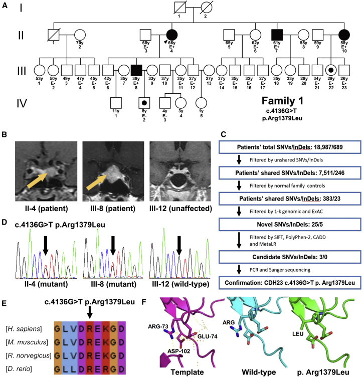
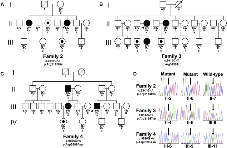
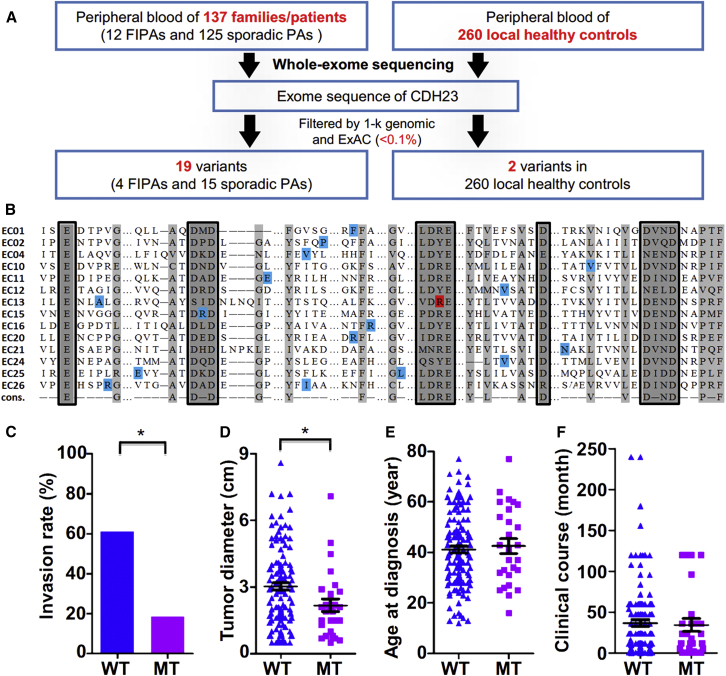
Pituitary adenoma (PA) is one of the most common intracranial neoplasms. Several genetic predisposing factors for PA have been identified, but they account for a small portion of cases. In this study, we sought to identify the PA genetic risk factors by focusing on causative mutations for PAs. Among the 4 affected and 17 asymptomatic members from one family with familial PA, whole-exome sequencing identified cosegregation of the PA phenotype with the heterozygous missense mutation c.4136G>T (p.Arg1379Leu) in cadherin-related 23 (CDH23). This mutation causes an amino acid substitution in the calcium-binding motif of the extracellular cadherin (EC) domains of CDH23 and is predicted to impair cell-cell adhesion. Genomic screening in a total of 12 families with familial PA (20 individuals), 125 individuals with sporadic PA, and 260 control individuals showed that 33% of the families with familial PA (4/12) and 12% of individuals with sporadic PA (15/125) harbored functional CDH23 variants. In contrast, 0.8% of the healthy control individuals (2/260) carried functional CDH23 variants. Gene-based analysis also revealed a significant association between CDH23 genotype and PA (p = 5.54 × 10⁻⁷). Moreover, PA individuals who did not harbor functional CDH23 variants displayed tumors that were larger in size (p = 0.005) and more invasive (p < 0.001). Therefore, mutations in CDH23 are linked with familial and sporadic PA and could play important roles in the pathogenesis of PA.
Keywords: CDH23; familial pituitary adenoma; mutation; pituitary adenoma; whole-exome sequencing.
Copyright © 2017 American Society of Human Genetics. Published by Elsevier Inc. All rights reserved.
Figures
Similar articles
-
Very low frequency of germline GPR101 genetic variation and no biallelic defects with AIP in a large cohort of patients with sporadic pituitary adenomas.Eur J Endocrinol. 2016 Apr;174(4):523-30. doi: 10.1530/EJE-15-1044. Epub 2016 Jan 20. Eur J Endocrinol. 2016. PMID: 26792934
-
Frequency of familial pituitary adenoma syndromes among patients with functioning pituitary adenomas in a reference outpatient clinic.J Endocrinol Invest. 2017 Dec;40(12):1381-1387. doi: 10.1007/s40618-017-0725-8. Epub 2017 Jul 8. J Endocrinol Invest. 2017. PMID: 28689311
-
Germline Variants in Sporadic Pituitary Adenomas.J Endocr Soc. 2024 Apr 24;8(6):bvae085. doi: 10.1210/jendso/bvae085. eCollection 2024 Apr 6. J Endocr Soc. 2024. PMID: 38745824 Free PMC article.
-
Genetic mutations in sporadic pituitary adenomas--what to screen for?Nat Rev Endocrinol. 2015 Jan;11(1):43-54. doi: 10.1038/nrendo.2014.181. Epub 2014 Oct 28. Nat Rev Endocrinol. 2015. PMID: 25350067 Review.
-
[AIP mutations in familial and sporadic pituitary adenomas: local experience and review of the literature].Endocrinol Nutr. 2009 Aug-Sep;56(7):369-77. doi: 10.1016/S1575-0922(09)72456-8. Endocrinol Nutr. 2009. PMID: 19883897 Review. Spanish.
Cited by
-
A Common Variant in the CDK8 Gene Is Associated with Sporadic Pituitary Adenomas in the Portuguese Population: A Case-Control Study.Int J Mol Sci. 2022 Oct 4;23(19):11749. doi: 10.3390/ijms231911749. Int J Mol Sci. 2022. PMID: 36233050 Free PMC article.
-
Case report: Identification of potential prognosis-related LAG3 overexpression and DICER1 mutation in pituitary carcinoma: two cases.Front Neurosci. 2023 Oct 12;17:1191596. doi: 10.3389/fnins.2023.1191596. eCollection 2023. Front Neurosci. 2023. PMID: 37901430 Free PMC article.
-
The Genomic Landscape of Sporadic Prolactinomas.Endocr Pathol. 2019 Dec;30(4):318-328. doi: 10.1007/s12022-019-09587-0. Endocr Pathol. 2019. PMID: 31473917
-
Differential Methylation Profile in Fragile X Syndrome-Prone Offspring Mice after in Utero Exposure to Lactobacillus Reuteri.Genes (Basel). 2022 Jul 22;13(8):1300. doi: 10.3390/genes13081300. Genes (Basel). 2022. PMID: 35893036 Free PMC article.
-
Genetic and Epigenetic Causes of Pituitary Adenomas.Front Endocrinol (Lausanne). 2021 Jan 26;11:596554. doi: 10.3389/fendo.2020.596554. eCollection 2020. Front Endocrinol (Lausanne). 2021. PMID: 33574795 Free PMC article. Review.
References
-
- Lecoq A.L., Kamenický P., Guiochon-Mantel A., Chanson P. Genetic mutations in sporadic pituitary adenomas--what to screen for? Nat. Rev. Endocrinol. 2015;11:43–54. - PubMed
-
- Saeger W., Lüdecke D.K., Buchfelder M., Fahlbusch R., Quabbe H.J., Petersenn S. Pathohistological classification of pituitary tumors: 10 years of experience with the German Pituitary Tumor Registry. Eur. J. Endocrinol. 2007;156:203–216. - PubMed
-
- Elston M.S., McDonald K.L., Clifton-Bligh R.J., Robinson B.G. Familial pituitary tumor syndromes. Nat. Rev. Endocrinol. 2009;5:453–461. - PubMed
-
- Caimari F., Korbonits M. Novel Genetic Causes of Pituitary Adenomas. Clin. Cancer Res. 2016;22:5030–5042. - PubMed
-
- Toledo R.A., Lourenço D.M., Jr., Toledo S.P. Familial isolated pituitary adenoma: evidence for genetic heterogeneity. Front. Horm. Res. 2010;38:77–86. - PubMed
MeSH terms
Substances
LinkOut - more resources
Full Text Sources
Other Literature Sources
Medical
Molecular Biology Databases
