Review
. 2017:147:1-73.
doi: 10.1016/bs.pmbts.2017.02.005. Epub 2017 Mar 22.

Biochemical and Biological Attributes of Matrix Metalloproteinases

Affiliations
Review

Biochemical and Biological Attributes of Matrix Metalloproteinases

Ning Cui et al. Prog Mol Biol Transl Sci. 2017.

Abstract

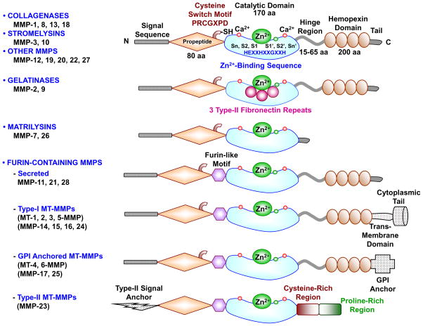
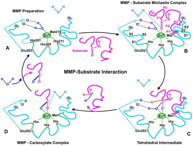
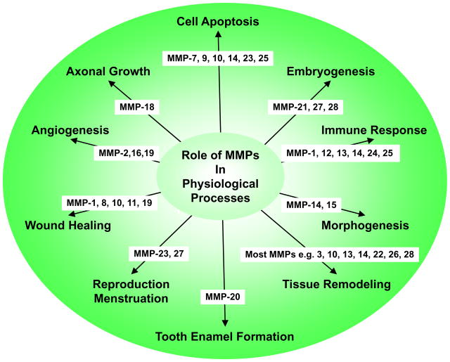
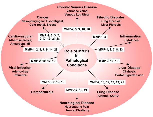
Matrix metalloproteinases (MMPs) are a family of zinc-dependent endopeptidases that are involved in the degradation of various proteins in the extracellular matrix (ECM). Typically, MMPs have a propeptide sequence, a catalytic metalloproteinase domain with catalytic zinc, a hinge region or linker peptide, and a hemopexin domain. MMPs are commonly classified on the basis of their substrates and the organization of their structural domains into collagenases, gelatinases, stromelysins, matrilysins, membrane-type (MT)-MMPs, and other MMPs. MMPs are secreted by many cells including fibroblasts, vascular smooth muscle (VSM), and leukocytes. MMPs are regulated at the level of mRNA expression and by activation of their latent zymogen form. MMPs are often secreted as inactive pro-MMP form which is cleaved to the active form by various proteinases including other MMPs. MMPs cause degradation of ECM proteins such as collagen and elastin, but could influence endothelial cell function as well as VSM cell migration, proliferation, Ca2+ signaling, and contraction. MMPs play a role in tissue remodeling during various physiological processes such as angiogenesis, embryogenesis, morphogenesis, and wound repair, as well as in pathological conditions such as myocardial infarction, fibrotic disorders, osteoarthritis, and cancer. Increases in specific MMPs could play a role in arterial remodeling, aneurysm formation, venous dilation, and lower extremity venous disorders. MMPs also play a major role in leukocyte infiltration and tissue inflammation. MMPs have been detected in cancer, and elevated MMP levels have been associated with tumor progression and invasiveness. MMPs can be regulated by endogenous tissue inhibitors of metalloproteinases (TIMPs), and the MMP/TIMP ratio often determines the extent of ECM protein degradation and tissue remodeling. MMPs have been proposed as biomarkers for numerous pathological conditions and are being examined as potential therapeutic targets in various cardiovascular and musculoskeletal disorders as well as cancer.

Keywords: Cell signaling; Collagen; Extracellular matrix; Protein degradation; Proteinases; Remodeling.


Conflict of interest statement

CONFLICT OF INTEREST

None

Figures

Fig. 1
Fig. 1
Major MMPs subtypes and their structure. A typical MMP consists of a propeptide, a catalytic metalloproteinase domain, a linker peptide (hinge region), and a hemopexin domain. The propeptide has a cysteine switch PRCGXPD whose cysteine sulfhydryl (–SH) group chelates the active site Zn2+, keeping the MMP in the latent proMMP zymogen form. The catalytic domain contains the Zn2+ binding motif HEXXHXXGXXH, two Zn2+ ions (one catalytic and one structural), specific S1, S2,…Sn and S1′, S2′,…Sn′ pockets, which confer specificity, and two or three Ca2+ ions for stabilization. Some MMPs show exceptions in their structures. Gelatinases have 3 type-II fibronectin repeats in the catalytic domain. Matrilysins have neither a hinge region nor a hemopexin domain. Furin-containing MMPs such as MMP-11, 21 and 28 have a furin-like pro-protein convertase recognition sequence in the propeptide C-terminus. MMP-28 has a slightly different cysteine switch motif PRCGVTD. Membrane-type MMPs (MT-MMPs) typically have a transmembrane domain and a cytosolic domain. MMP-17 and -25 have a glycosylphosphatidylinositol (GPI) anchor. MMP-23 lacks the consensus PRCGXPD motif, has a cysteine residue located in a different sequence ALCLLPA, may remain in the latent inactive proform through its type-II signal anchor, and has a cysteine-rich region and an immunoglobulin-like proline-rich region.
Fig. 2
Fig. 2
MMP-substrate interaction. MMP-3 is used as an example, and slight variations in the MMP-substrate-interaction and the positions of the conserved His and Glu may occur with other MMPs. Only the MMP catalytic domain is illustrated, and the remaining part of the MMP molecule is truncated by squiggles. A) In the quiescent MMP molecule, the catalytic Zn2+ is supported in the HEXXHXXGXXH-motif by binding to the imidazole rings of the 3 histidines His201, 205, 211. Additionally, the methionine-219 (Met219) in the conserved XBMX Met-turn acts as a hydrophobic base to further support the structure surrounding the catalytic Zn2+. In preparation of MMP for substrate binding, an incoming H2O molecule is polarized between the MMP acidic Zn2+ and basic glutamate-202 (Glu202). B) Using H+ from free H2O, the substrate carbonyl group binds to Zn2+, forming a Michaelis complex. This allows the MMP S1, S2, S3, …Sn pockets on the right side of Zn2+ and the primed S1′, S2′, S3′, …Sn′ pockets on the left side of Zn2+ to confer specific binding to the substrate P1, P2, P3, … Pn and the primed P1′, P2′, P3′, … Pn′ substituents, respectively. The MMP pockets are organized such that the S1 and S3 pockets are located away from the catalytic Zn2+, while the S2 pocket is closer to Zn2+. C) The substrate-bound H2O is freed, the Zn2+-bound O from the Glu-bound H2O executes a nucleophilic attack on the substrate carbon, and the Glu202 extracts a proton from the Glu-bound H2O to form an N-H bond with the substrate N, resulting in a tetrahedral intermediate. D) Freed H2O is taken up again, and the second proton from Glu-bound H2O is transferred to the substrate, forming an additional N-H bond. As a result, the substrate scissile C-N bond breaks, thus releasing the N portion of the substrate while the carboxylate portion of the substrate remains in an MMP-carboxylate complex. Another H2O is taken up, thus releasing the remaining carboxylate portion of the substrate, and the MMP is prepared to attack another substrate (A).
Fig. 3
Fig. 3
Representative roles of MMPs in physiological processes.
Fig. 4
Fig. 4
Representative roles of MMPs in pathological conditions. COPD, chronic obstructive pulmonary disease; MI, myocardial infarction

References

    1. Mittal R, Patel AP, Debs LH, Nguyen D, Patel K, Grati M, Mittal J, Yan D, Chapagain P, Liu XZ. Intricate Functions of Matrix Metalloproteinases in Physiological and Pathological Conditions. Journal of cellular physiology. 2016;231(12):2599–2621. - PubMed
    1. Kucukguven A, Khalil RA. Matrix metalloproteinases as potential targets in the venous dilation associated with varicose veins. Curr Drug Targets. 2013;14(3):287–324. - PMC - PubMed
    1. MacColl E, Khalil RA. Matrix Metalloproteinases as Regulators of Vein Structure and Function: Implications in Chronic Venous Disease. The Journal of pharmacology and experimental therapeutics. 2015;355(3):410–428. - PMC - PubMed
    1. Raffetto JD, Khalil RA. Matrix metalloproteinases and their inhibitors in vascular remodeling and vascular disease. Biochemical pharmacology. 2008;75(2):346–359. - PMC - PubMed
    1. Gross J, Lapiere CM. Collagenolytic activity in amphibian tissues: a tissue culture assay. Proc Natl Acad Sci U S A. 1962;48:1014–1022. - PMC - PubMed

Publication types

Substances