

Exposure of the Amino Terminus of Tau Is a Pathological Event in Multiple Tauopathies
- PMID: 28413156
- PMCID: PMC5818634
- DOI: 10.1016/j.ajpath.2017.01.019
Exposure of the Amino Terminus of Tau Is a Pathological Event in Multiple Tauopathies
Abstract
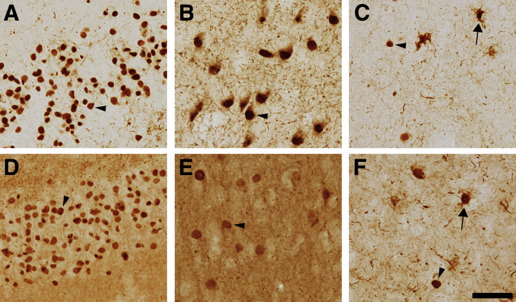
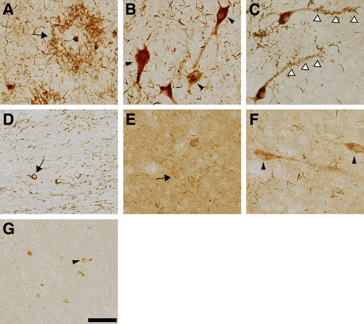
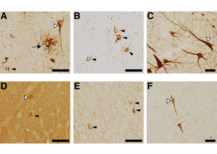
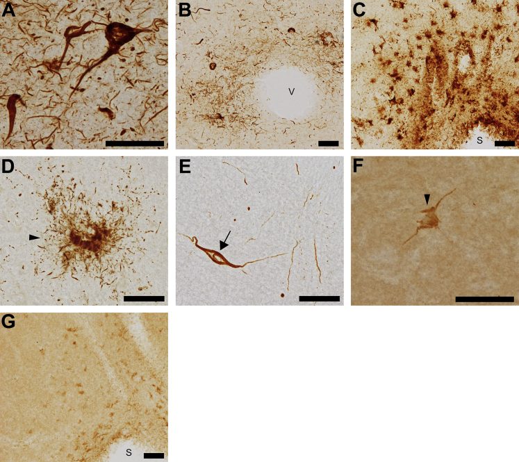
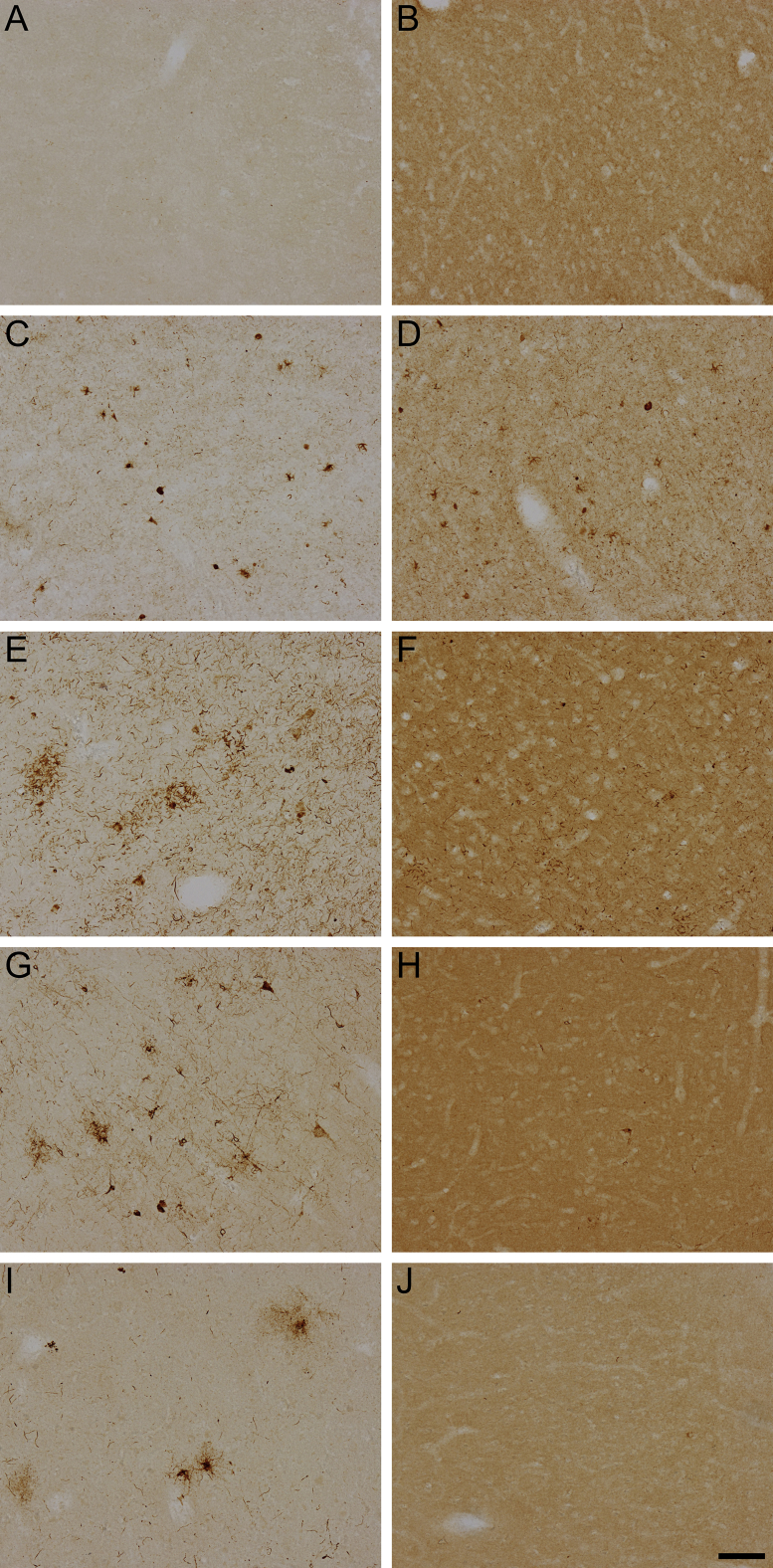
Pathological changes to the tau protein, including conformational changes and aggregation, are major hallmarks of a group of neurodegenerative disorders known as tauopathies. Among the conformational changes are alterations involving the extreme amino terminus of the protein, known as the phosphatase-activating domain (PAD). Aberrant PAD exposure induces a signaling cascade that leads to disruption of axonal transport, a critical function for neuronal survival. Conformational display of PAD is an early marker of pathological tau in Alzheimer disease (AD), but its role in other tauopathies has yet to be firmly established. We used a relatively novel N-terminal, conformation-sensitive antibody, TNT2, to determine whether misfolding in the amino terminus (ie, PAD exposure) occurs in non-AD tauopathies. We found that TNT2 specifically labeled pathological tau in post-mortem human brain tissue from Pick disease, progressive supranuclear palsy, corticobasal degeneration, and chronic traumatic encephalopathy, but did not label nonpathological, parenchymal tau. Tau13, another N-terminal antibody, was not sensitive to pathological N-terminal conformations. Tau13 did not readily distinguish between normal (ie, parenchymal tau) and pathological tau species and showed a range of effectiveness at identifying tau pathologies in the non-AD tauopathies. These findings demonstrate that the conformational display of the PAD in tau represents a common pathological event in many tauopathies.
Copyright © 2017 American Society for Investigative Pathology. Published by Elsevier Inc. All rights reserved.
Figures
Similar articles
-
Pathological conformations involving the amino terminus of tau occur early in Alzheimer's disease and are differentially detected by monoclonal antibodies.Neurobiol Dis. 2016 Oct;94:18-31. doi: 10.1016/j.nbd.2016.05.016. Epub 2016 May 31. Neurobiol Dis. 2016. PMID: 27260838 Free PMC article.
-
Detection of Alzheimer Disease (AD)-Specific Tau Pathology in AD and NonAD Tauopathies by Immunohistochemistry With Novel Conformation-Selective Tau Antibodies.J Neuropathol Exp Neurol. 2018 Mar 1;77(3):216-228. doi: 10.1093/jnen/nly010. J Neuropathol Exp Neurol. 2018. PMID: 29415231 Free PMC article.
-
Selective tau tyrosine nitration in non-AD tauopathies.Acta Neuropathol. 2012 Jan;123(1):119-32. doi: 10.1007/s00401-011-0898-8. Epub 2011 Nov 6. Acta Neuropathol. 2012. PMID: 22057784 Free PMC article.
-
Tauopathies as clinicopathological entities.Parkinsonism Relat Disord. 2016 Jan;22 Suppl 1(0 1):S29-33. doi: 10.1016/j.parkreldis.2015.09.020. Epub 2015 Sep 8. Parkinsonism Relat Disord. 2016. PMID: 26382841 Free PMC article. Review.
-
[Neuropathology of tauopathy].Brain Nerve. 2013 Dec;65(12):1445-58. Brain Nerve. 2013. PMID: 24323931 Review. Japanese.
Cited by
-
Phosphomimetics at Ser199/Ser202/Thr205 in Tau Impairs Axonal Transport in Rat Hippocampal Neurons.Mol Neurobiol. 2023 Jun;60(6):3423-3438. doi: 10.1007/s12035-023-03281-3. Epub 2023 Mar 2. Mol Neurobiol. 2023. PMID: 36859689 Free PMC article.
-
RNA binding proteins co-localize with small tau inclusions in tauopathy.Acta Neuropathol Commun. 2018 Aug 1;6(1):71. doi: 10.1186/s40478-018-0574-5. Acta Neuropathol Commun. 2018. PMID: 30068389 Free PMC article.
-
Production of recombinant tau oligomers in vitro.Methods Cell Biol. 2017;141:45-64. doi: 10.1016/bs.mcb.2017.06.005. Epub 2017 Jul 14. Methods Cell Biol. 2017. PMID: 28882311 Free PMC article.
-
Frontotemporal Lobar Dementia Mutant Tau Impairs Axonal Transport through a Protein Phosphatase 1γ-Dependent Mechanism.J Neurosci. 2021 Nov 10;41(45):9431-9451. doi: 10.1523/JNEUROSCI.1914-20.2021. Epub 2021 Oct 4. J Neurosci. 2021. PMID: 34607969 Free PMC article.
-
Passive immunotherapy for N-truncated tau ameliorates the cognitive deficits in two mouse Alzheimer's disease models.Brain Commun. 2020 Apr 6;2(1):fcaa039. doi: 10.1093/braincomms/fcaa039. eCollection 2020. Brain Commun. 2020. PMID: 32954296 Free PMC article.
References
-
- Kovacs G.G. Invited review: neuropathology of tauopathies: principles and practice. Neuropathol Appl Neurobiol. 2015;41:3–23. - PubMed
-
- Braak H., Braak E. Neuropathological stageing of Alzheimer-related changes. Acta Neuropathol. 1991;82:239–259. - PubMed
-
- Braak E., Braak H., Mandelkow E.M. A sequence of cytoskeleton changes related to the formation of neurofibrillary tangles and neuropil threads. Acta Neuropathol. 1994;87:554–567. - PubMed
-
- Iwatsubo T., Hasegawa M., Ihara Y. Neuronal and glial tau-positive inclusions in diverse neurologic diseases share common phosphorylation characteristics. Acta Neuropathol. 1994;88:129–136. - PubMed
MeSH terms
Substances
Grants and funding
LinkOut - more resources
Full Text Sources
Other Literature Sources
