. 2009 Feb 23;21(8):847-867.
doi: 10.1002/adma.200801478. Epub 2008 Dec 4.

Polymeric Materials for Gene Delivery and DNA Vaccination

Affiliations

Polymeric Materials for Gene Delivery and DNA Vaccination

David N Nguyen et al. Adv Mater. .

Abstract

Gene delivery holds great potential for the treatment of many different diseases. Vaccination with DNA holds particular promise, and may provide a solution to many technical challenges that hinder traditional vaccine systems including rapid development and production and induction of robust cell-mediated immune responses. However, few candidate DNA vaccines have progressed past preclinical development and none have been approved for human use. This Review focuses on the recent progress and challenges facing materials design for nonviral DNA vaccine drug delivery systems. In particular, we highlight work on new polymeric materials and their effects on protective immune activation, gene delivery, and current efforts to optimize polymeric delivery systems for DNA vaccination.


Figures

Figure 1
Figure 1
Simplified pathways of T-cell activation by dendritic cells leading to cell-mediated and humoral immune responses to DNA vaccines. Direct transfection of the antigen presenting cell (APC), here a dendritic cell, leads to intracellular production of antigen and presentation on MHCI to CD8+T-cells. Antigen can also be released by another transfected cell, and APC uptake of extracellular antigen leads to MHCII presentation to CD4+ T-cells. Class switching allows diversion of antigen from one presentation pathway to the other. Costimulatory signals are also required for T-cell activation. CD8+ T-cells expand and differentiate into cytotoxic T-cells (CTL) to kill infected cells and tumors. CD4+ T-cells expand and differentiate into various types of helper T-cells (TH) that activate B-cells to differentiate into plasma B cells and produce high affinity antibodies for opsonization or activation of antibody-dependent cell-mediated cytotoxicity (ADCC). CD4+ TH cells stimulate CTL responses as well.
Figure 2
Figure 2
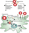
Cellular barriers to antigen presentation. Vector-nucleic acid particles (VNP) associate with the APC and enter through a variety of pathways[1] that are directed for degradation in the late endosome/phagosome.[2] The VNP must protect the plasmid DNA[3] and provide a method to escape the endosome.[4] The VNP then disassociates plasmid DNA[5] and may facilitate nuclear uptake[6] leading to gene expression.[7] Antigen encoded by the plasmid DNA is then processed and presented on MHC for immune responses[8] (see Table 1). Receptor–ligand interactions commonly found on APCs include: mannose and the mannose receptor (MR), antibody-coated particles and Fc receptors such as Fc-gamma receptor (FCγR) that recognizes IgG, complement-coated particles and the complement receptors (CR), and other scavenger receptors (SR).
Figure 3
Figure 3
Schematic of common microparticle and nanoparticle strategies for DNA vaccine delivery. a) Microparticles encapsulating supercoiled plasmid DNA in the matrix. b) Micro-particles co-encapsulating plasmid DNA and a secondary transfection agent. c) Cationic microparticles with surface adsorbed DNA plasmids. d) Nanoparticle electrostatic complexes of a cationic transfection agent and anionic plasmid DNA. e) Nanoparticles incorporating a targeting ligand to increase APC association and uptake.
Figure 4
Figure 4
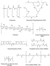
Polymers commonly used as nonviral gene delivery vectors.
Figure 5
Figure 5
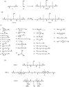
Structures and schemes for end-modification of PBAEs. a) Synthesis of acrylate-terminated C32 (C32-Ac). b) End-capping of C32-Ac. c) Amine-containing monomers used for end-capping, d) Other acrylate-terminated PBAEs that were end-modified.
Figure 6
Figure 6
Gene delivery efficacy of end-modified PBAE/DNA particles a) as measured in the COS-7 (blue bars) and HepG2 (red bars) cell lines. Optimized, unmodified 32-terminated C32 is shown at a low 20:1 polymer/DNA weight ratio (C32L) and high 100:1 ratio (C32H). b) C32-103 tested head-to-head with adenovirus at a multiplicity of infection (MOI) of 100 in HUVECs, primary human endothelial cells. c) Other top PBAEs compared to leading nonviral transfection agents PEI and Lipofectamine 2000 and to adenovirus at a range of MOIs. Reproduced with permission from [140].
Figure 7
Figure 7
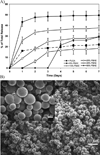
PBAE-modified PLGA microspheres. a) Release of plasmid DNA from PLGA microspheres incorporating up to 50% PBAE (w/w). 100% PLGA (0% PBAE) shown in dashed lines. Increasing the PBAE content reduces total DNA release and suppresses burst release. b) SEM image of PLGA microspheres containing 25% PBAE. Reproduced with permission from [182] Copyright 2005 Elsevier.
Figure 8
Figure 8
Tumor suppression following immunization with PLGA-PBAE microspheres. Antigen-specific, CTL-mediated suppression of SIY-bearing target tumors (right) compared to SIY-free non-target tumors (left) following intradermal (ID) vaccination with PLGA–PBAE microspheres that deliver plasmid DNA encoding for the SIY antigen. Tumor size (2-D) was measured at days 7 (blue), 9 (red), 11 (orange), and 15 (pink) after subcutaneous tumor injection; * denotes one mouse with complete regression of targeted tumors. Reproduced with permission from Ref.[109]. Copyright 2004 The National Academy of Sciences of the United States of America.
Figure 9
Figure 9
POE DNA release kinetics. Release of plasmid DNA from POE1 (circles) and POE2 (triangles) at pH 7 (empty shapes) and after switch to pH 5.4 (filled shapes) indicated by the arrow. POE microspheres exhibit classic two-phase release kinetics with an initial burst phase followed by a slow continuous phase as pH 7, or a rapid acid-catalyzed release at low pH such as that experienced within the phagosome. Reproduced with permission from Ref.[161]. Copyright 2004 Nature Publishing Group.
Figure 10
Figure 10
Tumor suppression following immunization with POE microspheres. Antigen-specific, CTL-mediated suppression of SIY-bearing target tumors (left) compared to SIY-free non-target tumors (right) following intradermal vaccination with POE microspheres that deliver plasmid DNA encoding for the SIY antigen. * denotes p < 0.05 compared to naked DNA (ID); # denotes p < 0.03 compared to PLGA microspheres. Reproduced with permission from [161]. Copyright 2004 Nature Publishing Group.
Figure 11
Figure 11
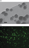
Nanoparticles of end-modified PBAE. a) TEM image of PBAE/DNA nanoparticles. Scale bar is 100 nm. b) Transfection of a confluent monolayer of the DC2.4 murine dendritic cell line with PBAE/DNA nanoparticles encoding green fluorescent protein (transfection efficiency ~20%).
Figure 12
Figure 12
PAMAM–PEG–mannose linear-dendritic block copolymers, a) Schematic structure and functionality of PAMAM–PEG–mannose linear-dendritic block copolymers complexed with plasmid DNA. b) Third generation exhibiting chemical structure and functionality. c) TEM image of sixth generation complexed with plasmid DNA. Reproduced with permission from [200].

References

    1. List of Vaccine-Preventable Diseases, Centers for Disease Control and Prevention (CDC) [accessed 19 November, 2008]; http://www.cdc.gov/vaccines/vpd-vac/vpd-list.htm.
    1. Robinson HL. Clin. Pharmacol. Ther. 2007;82:686. - PubMed
    1. Berzofsky JA, Ahlers JD, Belyakov IM. Nat. Rev. Immunol. 2001;1:209. - PubMed
    1. Guermonprez P, Valladeau J, Zitvogel L, Thery C, Amigorena S. Annu. Rev. Immunol. 2002;20:621. - PubMed
    1. Sharma AK, Khuller GK. Immunol. Cell Biol. 2001;79:537. - PubMed

LinkOut - more resources