

Case Reports
doi: 10.1159/000458516.
eCollection 2017 Jan-Apr.
Choroidal Nevus-Associated Neovascular Membrane Demonstrated by OCT Angiography
Affiliations
- PMID: 28413408
- PMCID: PMC5346919
- DOI: 10.1159/000458516
Item in Clipboard
Case Reports
Choroidal Nevus-Associated Neovascular Membrane Demonstrated by OCT Angiography
Case Rep Ophthalmol.
.
Abstract
We present a case of choroidal nevus, complicated by a choroidal neovascular membrane (CNV) that was detected by OCT angiography. Choroidal nevi are relatively common intraocular tumors. The presence of subretinal and intraretinal fluids can indicate that a CNV has occurred as a complication, warranting prompt management. However, subretinal and intraretinal fluids are also documented in nevi without CNV. OCT angiography may be of great help in determining whether those fluids are associated or not with a CNV, therefore guiding therapy.
Keywords: Choroidal neovascular membrane; Choroidal nevus; OCT angiography; Swept-source OCT.
Figures
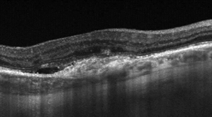
Swept-source OCT B-scan. This averaged picture at the fovea reveals disorganization of the outer retinal layers above the nevus as well as subretinal fluid. The borders of the nevus can be precisely delineated.

OCT angiography. Clinical examination and B-scan OCT cannot rule out the presence of a CNV. OCT angiography demonstrates a vascular lesion characterized by branching capillaries anastomosing into larger vessels and surrounded by a hypointense halo.

Fluorescein angiography reveals a classic CNV within the nevus.
References
-
- Shields CL, Pellegrini M, Ferenczy SR, Shields JA. Enhanced depth imaging optical coherence tomography of intraocular tumors: from placid to seasick to rock and rolling topography – the 2013 Francesco Orzalesi Lecture. Retina. 2014;34:1495–1512. - PubMed
-
- Francis JH, Pang CE, Abramson DH, Milman T, Folberg R, Mrejen S, Freund KB. Swept-source optical coherence tomography features of choroidal nevi. Am J Ophthalmol. 2015;159:169–176. e161. - PubMed
-
- Shields CL, Furuta M, Mashayekhi A, Berman EL, Zahler JD, Hoberman DM, Dinh DH, Shields JA. Clinical spectrum of choroidal nevi based on age at presentation in 3,422 consecutive eyes. Ophthalmology. 2008;115:546–552. e542. - PubMed
-
- Chiang A, Bianciotto C, Maguire JI, Park CH, Baker PS, Shields JA, Shields CL. Intravitreal bevacizumab for choroidal neovascularization associated with choroidal nevus. Retina. 2012;32:60–67. - PubMed
-
- Papastefanou VP, Nogueira V, Hay G, Andrews RM, Harris M, Cohen VM, Sagoo MS. Choroidal naevi complicated by choroidal neovascular membrane and outer retinal tubulation. Br J Ophthalmol. 2013;97:1014–1019. - PubMed
Publication types
LinkOut - more resources
Full Text Sources
Other Literature Sources
