. 2017 Mar 8;7(4):e00640.
doi: 10.1002/brb3.640. eCollection 2017 Apr.

White matter connections of the inferior parietal lobule: A study of surgical anatomy

Affiliations

White matter connections of the inferior parietal lobule: A study of surgical anatomy

Joshua D Burks et al. Brain Behav. .

Abstract

Introduction: Interest in the function of the inferior parietal lobule (IPL) has resulted in increased understanding of its involvement in visuospatial and cognitive functioning, and its role in semantic networks. A basic understanding of the nuanced white-matter anatomy in this region may be useful in improving outcomes when operating in this region of the brain. We sought to derive the surgical relationship between the IPL and underlying major white-matter bundles by characterizing macroscopic connectivity.

Methods: Data of 10 healthy adult controls from the Human Connectome Project were used for tractography analysis. All IPL connections were mapped in both hemispheres, and distances were recorded between cortical landmarks and major tracts. Ten postmortem dissections were then performed using a modified Klingler technique to serve as ground truth.

Results: We identified three major types of connections of the IPL. (1) Short association fibers connect the supramarginal and angular gyri, and connect both of these gyri to the superior parietal lobule. (2) Fiber bundles from the IPL connect to the frontal lobe by joining the superior longitudinal fasciculus near the termination of the Sylvian fissure. (3) Fiber bundles from the IPL connect to the temporal lobe by joining the middle longitudinal fasciculus just inferior to the margin of the superior temporal sulcus.

Conclusions: We present a summary of the relevant anatomy of the IPL as part of a larger effort to understand the anatomic connections of related networks. This study highlights the principle white-matter pathways and highlights key underlying connections.

Keywords: DTI; anatomy; angular; gyrus; inferior parietal; supramarginal; tractography; white matter.


Figures

Figure 1
Figure 1
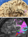
Superficial anatomy of the inferior parietal lobule (IPL). (a) The supramarginal gyrus (SMG) loops around the postero‐superior extension of the lateral sulcus (i.e., the Sylvian fissure). The angular gyrus (AG) similarly wraps around at the parietal extension of the STS. (b) Fiber dissection revealing the superficial connections of the SMG and AG. These tracts primarily facilitate local and intragyral connectivity, taking an oblique angle from the cortical surface. Some fibers from the SMG and AG travel superiorly to the superior parietal lobule (SPL). (c, d) Tractography demonstrating the orientation of superficial fibers from the IPL. Lateral (c) and supero‐lateral (d) views illustrate the fronto‐occipital and temporo‐occipital orientations of fibers connecting to the SMG, and the temporo‐occipital orientation of fibers connecting to the AG. Pink: SMG, fibers connecting to the SMG; Blue: AG, fibers connecting to the AG; IPS, intraparietal sulcus; LS, lateral sulcus (or Sylvian fissure); PN, preoccipital notch; STS, superior temporal sulcus
Figure 2
Figure 2
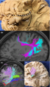
Short association fibers of the parietal lobe. Short association fibers of the cerebrum have a variable morphology, but tend to be “U‐shaped” as they connect adjacent gyri, as illustrated by (a). Short association fibers of the superior parietal lobule are shown with tractography (b) and gross dissection (c). Short association fibers of the supramarginal gyrus (SMG) have a distinct morphology (d), as its short association fibers must negotiate the lateral sulcus. As fiber bundles leave the major white‐matter pathways to connect to the cortical surface, they become intertwined and twist slightly as they course superficially. The hydra‐like shape of these fibers is illustrated by tractography showing isolated fibers in (e) and fibers of the SMG in total (f). Gross dissection of association fibers beneath the cortex of the SMG is shown in (g). LS, lateral sulcus; SLF, superior longitudinal fasciculus
Figure 3
Figure 3
The relationship between the inferior parietal lobule (IPL) and the insula. Gross dissection (a) and tractography (b) illustrate the proximity of the insula to fibers connecting the IPL to the frontal lobe. Fibers connecting to frontal regions and the AG curve with the arcuate fasciculus around the posterior edge of the insula before becoming part of the superior longitudinal fasciculus (SLF), passing directly superior to the insula. Similarly, fibers connecting to frontal regions and the SMG join the SLF to course antero‐posteriorly just above the insula. Red, inferior longitudinal fasciculus; Blue, inferior fronto‐occipital fasciculus; Green, middle longitudinal fasciculus; Purple, SMG/fibers connecting to the SMG; Light Blue, AG /fibers connecting to the AG; white, superior longitudinal fasciculus; CS, central sulcus; ITG, inferior temporal gyrus; LS, lateral sulcus; MFG, middle frontal gyrus; PO, pars orbitalis; SFG, superior frontal gyrus; SLF/AG, superior longitudinal fasciculus/arcuate fasciculus; SMG, supramarginal gyrus
Figure 4
Figure 4
White matter anatomy of the inferior parietal lobule (IPL). The AF courses beneath the SMG around the termination of the Sylvian fissure to connect frontal areas to the posterior MTG and STG as shown in (a) and (b). The SMG connects with frontal areas by coursing with SLF III, and the AG connects with frontal areas via SLF II, also shown in (b). Some fibers from the SMG and AG travel a short distance to the temporal lobes with the AF. Once in the temporal lobe, these fibers travel parallel to the MdLF (b and c). The MdLF is shown connecting the STG to the cuneus in (d), passing deep to the IPL and AF. AF, arcuate fasciculus; AG, angular gyrus; C, cuneus; IFG, inferior frontal gyrus; IFOF, inferior fronto‐occipital fasciculus; ILF, inferior longitudinal fasciculus; L, lingual gyrus; LS, lateral sulcus; MdLF, middle longitudinal fasciculus; MTG, middle temporal gyrus; SLF, superior longitudinal fasciculus; SMG, supramarginal gyrus; STG, superior temporal gyrus
Figure 5
Figure 5
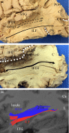
White‐matter pathways deep to the inferior parietal and posterior temporal lobes. Running medial and inferior to the fibers of the middle longitudinal fasciculus (MdLF) are the bundles of the inferior longitudinal fasciculus (ILF), which travel with fibers connecting the inferior parietal lobule to the ITG. The ILF is shown by fiber dissection in (a). The IFOF runs from prefrontal regions in the frontal lobe into the medial temporal lobe inferior to the insula, before coursing medial and parallel to the MdLF and terminating throughout the occipital lobe, as shown in (b). These tracts are shown by tractography in (c). Blue, IFOF; Red, ILF; Cu, cuneus; CF, calcarine fissure; IFG, inferior frontal gyrus; IFOF, inferior fronto‐occipital fasciculus; ITG, inferior temporal gyrus; L, lingual gyrus; LS, lateral sulcus; PN, preoccipital notch; POS, parieto‐occipital sulcus; STG, superior temporal gyrus; MdLF, middle longitudinal fasciculus
Figure 6
Figure 6
Defining D1 and D2. T1‐weighted coronal MR imaging illustrating D1, which was the shortest distance between the termination of the Sylvian fissure and the superior longitudinal fasciculus, is shown in (a). A slice taken slightly posterior to (a), illustrating D2, which was the shortest distance between the termination of the superior temporal sulcus and the arcuate fasciculus, is shown in (b). Both distances were measured in imaging and anatomical specimens. White: superior longitudinal fasciculus/arcuate fasciculus; Red: distance measured

Similar articles

Cited by

References

    1. Alain, C. , He, Y. , & Grady, C. (2008). The contribution of the inferior parietal lobe to auditory spatial working memory. Journal of Cognitive Neuroscience, 20, 285–295. - PubMed
    1. Almeida, J. , Mahon, B. Z. , Nakayama, K. , & Caramazza, A. (2008). Unconscious processing dissociates along categorical lines. Proceedings of the National Academy of Sciences of the United States of America, 105, 15214–15218. - PMC - PubMed
    1. Averbeck, B. B. , Battaglia‐Mayer, A. , Guglielmo, C. , & Caminiti, R. (2009). Statistical analysis of parieto‐frontal cognitive‐motor networks. Journal of Neurophysiology, 102, 1911–1920. - PubMed
    1. Binder, J. R. (2015). The Wernicke area: Modern evidence and a reinterpretation. Neurology, 85, 2170–2175. - PMC - PubMed
    1. Binder, J. R. , & Desai, R. H. (2011). The neurobiology of semantic memory. Trends in Cognitive Sciences, 15, 527–536. - PMC - PubMed

LinkOut - more resources